 Movie
Movie Controller


+ Open data

- Basic information
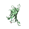
| Entry | Database: PDB / ID: 1sle | ||||||
|---|---|---|---|---|---|---|---|
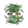
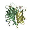
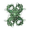
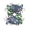
| Title | STREPTAVIDIN, PH 5.0, BOUND TO CYCLIC PEPTIDE AC-CHPQGPPC-NH2 | ||||||
 Components Components |
| ||||||
 Keywords Keywords | COMPLEX(BIOTIN-BINDING PROTEIN/PEPTIDE) / COMPLEX(BIOTIN-BINDING PROTEIN-PEPTIDE) / COMPLEX(BIOTIN-BINDING PROTEIN-PEPTIDE) complex | ||||||
| Function / homology |  Function and homology information Function and homology information | ||||||
| Biological species |  Streptomyces avidinii (bacteria) Streptomyces avidinii (bacteria) | ||||||
| Method |  X-RAY DIFFRACTION / Resolution: 2 Å X-RAY DIFFRACTION / Resolution: 2 Å | ||||||
 Authors Authors | Katz, B.A. | ||||||
 Citation Citation | Journal: Biochemistry / Year: 1995 Title: Binding to protein targets of peptidic leads discovered by phage display: crystal structures of streptavidin-bound linear and cyclic peptide ligands containing the HPQ sequence Authors: Katz, B.A. | ||||||
| History |
|
- Structure visualization
| Structure viewer | Molecule: MolmilJmol/JSmol |
|---|
- Downloads & links
-Download
| PDBx/mmCIF format | 1sle.cif.gz | 77.6 KB | Display | PDBx/mmCIF format |
|---|---|---|---|---|
| PDB format | pdb1sle.ent.gz | 58.8 KB | Display | PDB format |
| PDBx/mmJSON format | 1sle.json.gz | Tree view | PDBx/mmJSON format | |
| Others |  Other downloads Other downloads |
-Validation report
| Arichive directory | https://data.pdbj.org/pub/pdb/validation_reports/sl/1sleftp://data.pdbj.org/pub/pdb/validation_reports/sl/1sle | HTTPS FTP |
|---|
-Related structure data


-Links
 PDBj
PDBj- Assembly
| Deposited unit | 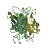
| ||||||||
|---|---|---|---|---|---|---|---|---|---|
| 1 | 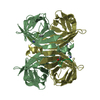
| ||||||||
| Unit cell |
| ||||||||
| Noncrystallographic symmetry (NCS) | NCS oper: (Code: given Matrix: (-1, -0.029, -0.007), Vector: Details | MTRIX THE TRANSFORMATIONS PRESENTED ON MTRIX RECORDS BELOW DESCRIBE NON-CRYSTALLOGRAPHIC RELATIONSHIPS AMONG THE VARIOUS DOMAINS IN THIS ENTRY. APPLYING THE APPROPRIATE MTRIX TRANSFORMATION TO THE RESIDUES LISTED FIRST WILL YIELD APPROXIMATE COORDINATES FOR THE RESIDUES LISTED SECOND. APPLIED TO TRANSFORMED TO MTRIX RESIDUES RESIDUES RMSD M1 B 13 .. M 8 D 13 .. P 8 0.646 SYMMETRY THE CRYSTALLOGRAPHIC SYMMETRY TRANSFORMATIONS PRESENTED BELOW GENERATE THE SUBUNITS OF THE POLYMERIC MOLECULE. STREPTAVIDIN IS A TETRAMERIC PROTEIN. THE CRYSTALLOGRAPHIC TRANSFORMATION GIVEN HERE GENERATES THE TETRAMER FROM THE DIMER FOUND IN THE ASYMMETRIC UNIT OF THE CRYSTALS. APPLIED TO RESIDUES: B 13 .. B 133 D 13 .. D 133 M 2 .. M 6 P 1 .. P 6 SYMMETRY1 1 1.000000 0.000000 0.000000 0.00000 SYMMETRY2 1 0.000000 -1.000000 0.000000 0.00000 SYMMETRY3 1 0.000000 0.000000 -1.000000 0.00000 | |
-Components
| #1: Protein | Mass: 14181.324 Da / Num. of mol.: 2 / Source method: isolated from a natural source / Source: (natural) Streptomyces avidinii (bacteria) / References: UniProt: P22629#2: Protein/peptide | Mass: 863.018 Da / Num. of mol.: 2 / Source method: isolated from a natural source #3: Water | ChemComp-HOH / |  Mass: 18.015 Da / Num. of mol.: 130 / Source method: isolated from a natural source / Formula: H2O Mass: 18.015 Da / Num. of mol.: 130 / Source method: isolated from a natural source / Formula: H2OHas protein modification | Y | |
|---|
-Experimental details
-Experiment
| Experiment | Method: X-RAY DIFFRACTION |
|---|
- Sample preparation
| Crystal | Density Matthews: 2.16 Å3/Da / Density % sol: 43.01 % |
|---|---|
| Crystal grow | pH: 5 / Details: pH 5.0 |
| Crystal | *PLUS Density % sol: 21 % |
| Crystal grow | *PLUS pH: 4 / Method: vapor diffusion, sitting drop |
| Components of the solutions | *PLUS Conc.: 15 mg/ml / Common name: peptide |
-Data collection
| Diffraction source | Wavelength: 1.5418 |
|---|---|
| Detector | Type: SIEMENS / Detector: AREA DETECTOR |
| Radiation | Monochromatic (M) / Laue (L): M / Scattering type: x-ray |
| Radiation wavelength | Wavelength: 1.5418 Å / Relative weight: 1 |
| Reflection | Num. obs: 16042 / % possible obs: 56 % / Observed criterion σ(I): 3.5 / Redundancy: 4 % / Rmerge(I) obs: 0.075 |
| Reflection | *PLUS Highest resolution: 1.9 Å / Lowest resolution: 50 Å / Num. measured all: 64474 / Rmerge(I) obs: 0.075 |
- Processing
| Software |
| ||||||||||||||||||||||||||||||||||||||||||||||||||||||||||||
|---|---|---|---|---|---|---|---|---|---|---|---|---|---|---|---|---|---|---|---|---|---|---|---|---|---|---|---|---|---|---|---|---|---|---|---|---|---|---|---|---|---|---|---|---|---|---|---|---|---|---|---|---|---|---|---|---|---|---|---|---|---|
| Refinement | Resolution: 2→7.5 Å / σ(F): 1 Details: CRYST1 CELL AXES CHOSEN TO CORRESPOND TO COORDINATES OF STREPTAVIDIN DEPOSITED BY WEBER ET AL. IN THE PDB (ENTRY 1PTS).
| ||||||||||||||||||||||||||||||||||||||||||||||||||||||||||||
| Displacement parameters | Biso mean: 22 Å2 | ||||||||||||||||||||||||||||||||||||||||||||||||||||||||||||
| Refinement step | Cycle: LAST / Resolution: 2→7.5 Å
| ||||||||||||||||||||||||||||||||||||||||||||||||||||||||||||
| Refine LS restraints |
| ||||||||||||||||||||||||||||||||||||||||||||||||||||||||||||
| Software | *PLUS Name: X-PLOR / Classification: refinement | ||||||||||||||||||||||||||||||||||||||||||||||||||||||||||||
| Refinement | *PLUS | ||||||||||||||||||||||||||||||||||||||||||||||||||||||||||||
| Solvent computation | *PLUS | ||||||||||||||||||||||||||||||||||||||||||||||||||||||||||||
| Displacement parameters | *PLUS | ||||||||||||||||||||||||||||||||||||||||||||||||||||||||||||
| Refine LS restraints | *PLUS
|
