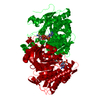
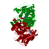
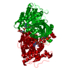
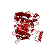
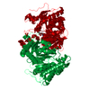
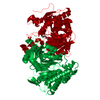
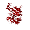
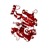
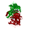
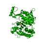
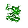
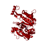
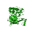
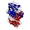
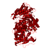
- PDB-4ajs: 3D structure of E. coli Isocitrate Dehydrogenase K100M mutant in ... -
+
Open data
ID or keywords:
Loading...
-
Basic information
Entry
Database: PDB / ID: 4ajs
Title
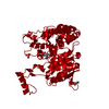
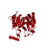
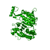
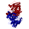
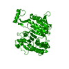
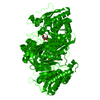
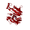
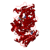
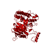
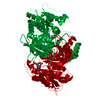
3D structure of E. coli Isocitrate Dehydrogenase K100M mutant in complex with isocitrate, magnesium(II), Adenosine 2',5'-biphosphate and ribosylnicotinamide-5'-phosphate
Components
ISOCITRATE DEHYDROGENASE [NADP]
Keywords
OXIDOREDUCTASE / OXIDATIVE BETA-DECARBOXYLATION
Function / homology
Function and homology information
isocitrate dehydrogenase (NADP+) / isocitrate dehydrogenase (NADP+) activity / glyoxylate cycle / guanosine tetraphosphate binding / tricarboxylic acid cycle / electron transport chain / NAD binding / response to oxidative stress / magnesium ion binding / cytoplasm / cytosol Similarity search - Function
Mass: 18.015 Da / Num. of mol.: 504 / Source method: isolated from a natural source / Formula: H2O
-
Details
Nonpolymer details
RIBOSYLNICOTINAMIDE-5'-PHOSPHATE (NMN): NADP FRAGMENT FROM HYDROLYSIS ADENOSINE 2',5'-BIPHOSPHATE ...RIBOSYLNICOTINAMIDE-5'-PHOSPHATE (NMN): NADP FRAGMENT FROM HYDROLYSIS ADENOSINE 2',5'-BIPHOSPHATE (A2P): NADP FRAGMENT FROM HYDROLYSIS
-
Experimental details
-
Experiment
Experiment
Method: X-RAY DIFFRACTION / Number of used crystals: 1
-
Sample preparation
Crystal
Density Matthews: 4.5 Å3/Da / Density % sol: 72 % / Description: NONE
Crystal grow
Details: 1.85 M NH4SO4, 50 MM CITRIC ACID/NA2HPO4 BUFFER PH 5.2, 0.1 M NACL AND 0.2 M DTT.
In the structure databanks used in Yorodumi, some data are registered as the other names, "COVID-19 virus" and "2019-nCoV". Here are the details of the virus and the list of structure data.
Jan 31, 2019. EMDB accession codes are about to change! (news from PDBe EMDB page)
EMDB accession codes are about to change! (news from PDBe EMDB page)
The allocation of 4 digits for EMDB accession codes will soon come to an end. Whilst these codes will remain in use, new EMDB accession codes will include an additional digit and will expand incrementally as the available range of codes is exhausted. The current 4-digit format prefixed with “EMD-” (i.e. EMD-XXXX) will advance to a 5-digit format (i.e. EMD-XXXXX), and so on. It is currently estimated that the 4-digit codes will be depleted around Spring 2019, at which point the 5-digit format will come into force.
The EM Navigator/Yorodumi systems omit the EMD- prefix.
Related info.:Q: What is EMD? / ID/Accession-code notation in Yorodumi/EM Navigator
Yorodumi is a browser for structure data from EMDB, PDB, SASBDB, etc.
This page is also the successor to EM Navigator detail page, and also detail information page/front-end page for Omokage search.
The word "yorodu" (or yorozu) is an old Japanese word meaning "ten thousand". "mi" (miru) is to see.
Related info.:EMDB / PDB / SASBDB / Comparison of 3 databanks / Yorodumi Search / Aug 31, 2016. New EM Navigator & Yorodumi / Yorodumi Papers / Jmol/JSmol / Function and homology information / Changes in new EM Navigator and Yorodumi
 Movie
Movie Controller

 Yorodumi
 Open data
 Basic information
 Components
 Keywords
 Function and homology information
 X-RAY DIFFRACTION /
  Authors
 Citation
 Structure visualization
 Downloads & links
 Other downloads
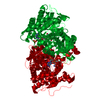


 PDBj
PDBj


 Assembly
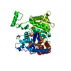


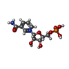


 Mass: 427.201 Da / Num. of mol.: 1 / Source method: obtained synthetically / Formula: C10H15N5O10P2
 Mass: 192.124 Da / Num. of mol.: 1 / Source method: obtained synthetically / Formula: C6H8O7
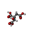
 Mass: 24.305 Da / Num. of mol.: 1 / Source method: obtained synthetically / Formula: Mg
 Mass: 335.227 Da / Num. of mol.: 1 / Source method: obtained synthetically / Formula: C11H16N2O8P
 Mass: 96.063 Da / Num. of mol.: 1 / Source method: obtained synthetically / Formula: SO4
 Sample preparation
 / Beamline: ID23-1 / Wavelength: 0.9795
  Processing