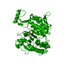
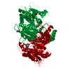
Mass: 45809.562 Da / Num. of mol.: 1 / Fragment: Full Length Source method: isolated from a genetically manipulated source Source: (gene. exp.) Escherichia coli (E. coli) / Gene: ICD OR ICDA OR ICDE OR B1136 / Production host: Escherichia coli (E. coli) / Keywords: wild type References: UniProt: P08200, isocitrate dehydrogenase (NADP+)
Mass: 18.015 Da / Num. of mol.: 240 / Source method: isolated from a natural source / Formula: H2O
-
Experimental details
-
Experiment
Experiment
Method: X-RAY DIFFRACTION / Number of used crystals: 1
-
Sample preparation
Crystal
Density Matthews: 4.37 Å3/Da / Density % sol: 71.83 %
Crystal grow
Temperature: 295 K / Method: vapor diffusion, hanging drop / pH: 7.5 Details: IDH, stored in metal-free final buffer, was diluted to 25, 30, 35 and 40 mg/ml using metal-free 2X buffer; (70mM Na2HPO4 , 18 mM citric acid, 200 mM NaCl, 0.4 mM DTT, pH 5.4). and was ...Details: IDH, stored in metal-free final buffer, was diluted to 25, 30, 35 and 40 mg/ml using metal-free 2X buffer; (70mM Na2HPO4 , 18 mM citric acid, 200 mM NaCl, 0.4 mM DTT, pH 5.4). and was crystallized from hanging drops using 5 l each of these IDH solutions and 5 l each of 34, 36, 38, 40, 42, and 44 % (NH4)2SO4 solutions in crystallization buffer (35mM Na2HPO4 , 9 mM citric acid, 100 mM NaCl, 0.2 mM DTT at pH 6.1, pH 7.5, VAPOR DIFFUSION, HANGING DROP, temperature 22K
Crystal grow
*PLUS
Method: unknown
-
Data collection
Diffraction
Mean temperature: 100 K
Diffraction source
Source: SYNCHROTRON / Site: ALS / Beamline: 5.0.2 / Wavelength: 1 Å
In the structure databanks used in Yorodumi, some data are registered as the other names, "COVID-19 virus" and "2019-nCoV". Here are the details of the virus and the list of structure data.
Jan 31, 2019. EMDB accession codes are about to change! (news from PDBe EMDB page)
EMDB accession codes are about to change! (news from PDBe EMDB page)
The allocation of 4 digits for EMDB accession codes will soon come to an end. Whilst these codes will remain in use, new EMDB accession codes will include an additional digit and will expand incrementally as the available range of codes is exhausted. The current 4-digit format prefixed with “EMD-” (i.e. EMD-XXXX) will advance to a 5-digit format (i.e. EMD-XXXXX), and so on. It is currently estimated that the 4-digit codes will be depleted around Spring 2019, at which point the 5-digit format will come into force.
The EM Navigator/Yorodumi systems omit the EMD- prefix.
Related info.:Q: What is EMD? / ID/Accession-code notation in Yorodumi/EM Navigator
Yorodumi is a browser for structure data from EMDB, PDB, SASBDB, etc.
This page is also the successor to EM Navigator detail page, and also detail information page/front-end page for Omokage search.
The word "yorodu" (or yorozu) is an old Japanese word meaning "ten thousand". "mi" (miru) is to see.
Related info.:EMDB / PDB / SASBDB / Comparison of 3 databanks / Yorodumi Search / Aug 31, 2016. New EM Navigator & Yorodumi / Yorodumi Papers / Jmol/JSmol / Function and homology information / Changes in new EM Navigator and Yorodumi
 Movie
Movie Controller

 Yorodumi
 Open data
 Basic information
 Components
 Keywords
 Function and homology information
 X-RAY DIFFRACTION /
  Authors
 Citation
 Structure visualization
 Downloads & links
 Other downloads
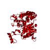


 PDBj
PDBj


 Assembly


 Mass: 96.063 Da / Num. of mol.: 1 / Source method: obtained synthetically / Formula: SO4
 Mass: 24.305 Da / Num. of mol.: 1 / Source method: obtained synthetically / Formula: Mg
 Mass: 192.124 Da / Num. of mol.: 1 / Source method: obtained synthetically / Formula: C6H8O7
 Mass: 92.094 Da / Num. of mol.: 3 / Source method: obtained synthetically / Formula: C3H8O3
 Sample preparation
 / Beamline: 5.0.2 / Wavelength: 1 Å
 Processing