 Movie
Movie Controller

[English] 日本語
 Yorodumi
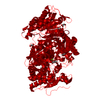
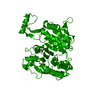
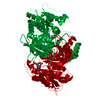
- PDB-1pb1: A four location model to explain the stereospecificity of proteins. -
+ Open data

- Basic information
| Entry | Database: PDB / ID: 1pb1 | ||||||
|---|---|---|---|---|---|---|---|
| Title | A four location model to explain the stereospecificity of proteins. | ||||||
 Components Components | Isocitrate dehydrogenase [NADP] | ||||||
 Keywords Keywords | OXIDOREDUCTASE / l-isocitrate / isocitrate dehydrogenase / enantiomer / specificity / stereospecificity | ||||||
| Function / homology |  Function and homology information Function and homology informationisocitrate dehydrogenase (NADP+) / isocitrate dehydrogenase (NADP+) activity / glyoxylate cycle / guanosine tetraphosphate binding / tricarboxylic acid cycle / electron transport chain / NAD binding / response to oxidative stress / magnesium ion binding / cytoplasm / cytosol Similarity search - Function | ||||||
| Biological species |  | ||||||
| Method |  X-RAY DIFFRACTION / SYNCHROTRON / MOLECULAR REPLACEMENT / Resolution: 1.7 Å X-RAY DIFFRACTION / SYNCHROTRON / MOLECULAR REPLACEMENT / Resolution: 1.7 Å | ||||||
 Authors Authors | Mesecar, A.D. / Koshland Jr., D.E. | ||||||
 Citation Citation | Journal: Nature / Year: 2000 Title: Structural Biology: A New Model for Protein Stereospecificity. Authors: Mesecar, A.D. / Koshland Jr., D.E. #1: Journal: IUBMB Life / Year: 2000Title: Sites of Binding and Orientation in a Four-Location Model for Protein Stereospecificity. Authors: Mesecar, A.D. / Koshland Jr., D.E. | ||||||
| History |
|
- Structure visualization
| Structure viewer | Molecule: MolmilJmol/JSmol |
|---|
- Downloads & links
-Download
| PDBx/mmCIF format | 1pb1.cif.gz | 101.9 KB | Display | PDBx/mmCIF format |
|---|---|---|---|---|
| PDB format | pdb1pb1.ent.gz | 77 KB | Display | PDB format |
| PDBx/mmJSON format | 1pb1.json.gz | Tree view | PDBx/mmJSON format | |
| Others |  Other downloads Other downloads |
-Validation report
| Arichive directory | https://data.pdbj.org/pub/pdb/validation_reports/pb/1pb1ftp://data.pdbj.org/pub/pdb/validation_reports/pb/1pb1 | HTTPS FTP |
|---|
-Related structure data


-Links
 PDBj
PDBj


- Assembly
| Deposited unit | 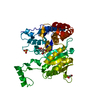
| ||||||||
|---|---|---|---|---|---|---|---|---|---|
| 1 | 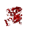
| ||||||||
| Unit cell |
|
-Components
| #1: Protein | Mass: 45809.562 Da / Num. of mol.: 1 Source method: isolated from a genetically manipulated source Details: wildtype / Source: (gene. exp.) References: UniProt: P08200, isocitrate dehydrogenase (NADP+) | ||
|---|---|---|---|
| #2: Chemical | ChemComp-SO4 /   Mass: 96.063 Da / Num. of mol.: 1 / Source method: obtained synthetically / Formula: SO4 Mass: 96.063 Da / Num. of mol.: 1 / Source method: obtained synthetically / Formula: SO4 | ||
| #3: Chemical | ChemComp-ICT / 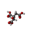  Mass: 192.124 Da / Num. of mol.: 1 / Source method: obtained synthetically / Formula: C6H8O7 Mass: 192.124 Da / Num. of mol.: 1 / Source method: obtained synthetically / Formula: C6H8O7 | ||
| #4: Chemical |   Mass: 92.094 Da / Num. of mol.: 3 / Source method: obtained synthetically / Formula: C3H8O3 Mass: 92.094 Da / Num. of mol.: 3 / Source method: obtained synthetically / Formula: C3H8O3#5: Water | ChemComp-HOH / |  Mass: 18.015 Da / Num. of mol.: 337 / Source method: isolated from a natural source / Formula: H2O Mass: 18.015 Da / Num. of mol.: 337 / Source method: isolated from a natural source / Formula: H2O |
-Experimental details
-Experiment
| Experiment | Method: X-RAY DIFFRACTION / Number of used crystals: 1 |
|---|
- Sample preparation
| Crystal | Density Matthews: 4.38 Å3/Da / Density % sol: 71.9 % |
|---|---|
| Crystal grow | Temperature: 295 K / Method: vapor diffusion, hanging drop / pH: 6.2 Details: IDH, stored in metal-free final buffer, was diluted to 25, 30, 35 and 40 mg/ml using metal-free 2X buffer; (70mM Na2HPO4 , 18 mM citric acid, 200 mM NaCl, 0.4 mM DTT, pH 5.4). and was ...Details: IDH, stored in metal-free final buffer, was diluted to 25, 30, 35 and 40 mg/ml using metal-free 2X buffer; (70mM Na2HPO4 , 18 mM citric acid, 200 mM NaCl, 0.4 mM DTT, pH 5.4). and was crystallized from hanging drops using 5 l each of these IDH solutions and 5 l each of 34, 36, 38, 40, 42, and 44 % (NH4)2SO4 solutions in crystallization buffer (35mM Na2HPO4 , 9 mM citric acid, 100 mM NaCl, 0.2 mM DTT at pH 5.4). , pH 6.2, VAPOR DIFFUSION, HANGING DROP, temperature 22K |
| Crystal grow | *PLUS Method: unknown |
-Data collection
| Diffraction | Mean temperature: 100 K |
|---|---|
| Diffraction source | Source: SYNCHROTRON / Site: ALS  / Beamline: 5.0.2 / Wavelength: 1 Å / Beamline: 5.0.2 / Wavelength: 1 Å |
| Detector | Type: ADSC QUANTUM 4 / Detector: CCD / Date: Jan 1, 1999 |
| Radiation | Protocol: SINGLE WAVELENGTH / Monochromatic (M) / Laue (L): M / Scattering type: x-ray |
| Radiation wavelength | Wavelength: 1 Å / Relative weight: 1 |
| Reflection | Resolution: 1.7→25 Å / Num. all: 91206 / Num. obs: 83266 / % possible obs: 92.7 % / Observed criterion σ(F): 0 / Observed criterion σ(I): 0 / Biso Wilson estimate: 17.7 Å2 |
| Reflection shell | Resolution: 1.7→1.76 Å / Rmerge(I) obs: 0.212 / Mean I/σ(I) obs: 6.8 / % possible all: 84.6 |
- Processing
| Software |
| ||||||||||||||||||||||||||||||||||||
|---|---|---|---|---|---|---|---|---|---|---|---|---|---|---|---|---|---|---|---|---|---|---|---|---|---|---|---|---|---|---|---|---|---|---|---|---|---|
| Refinement | Method to determine structure: MOLECULAR REPLACEMENT / Resolution: 1.7→24.79 Å / Rfactor Rfree error: 0.002 / Isotropic thermal model: RESTRAINED / Cross valid method: THROUGHOUT / σ(F): 0 / Stereochemistry target values: Engh & Huber
| ||||||||||||||||||||||||||||||||||||
| Solvent computation | Solvent model: FLAT MODEL / Bsol: 73.3181 Å2 / ksol: 0.398972 e/Å3 | ||||||||||||||||||||||||||||||||||||
| Displacement parameters | Biso mean: 21.4 Å2
| ||||||||||||||||||||||||||||||||||||
| Refine analyze |
| ||||||||||||||||||||||||||||||||||||
| Refinement step | Cycle: LAST / Resolution: 1.7→24.79 Å
| ||||||||||||||||||||||||||||||||||||
| Refine LS restraints |
| ||||||||||||||||||||||||||||||||||||
| LS refinement shell | Resolution: 1.7→1.76 Å / Rfactor Rfree error: 0.01 / Total num. of bins used: 10
| ||||||||||||||||||||||||||||||||||||
| Xplor file |
| ||||||||||||||||||||||||||||||||||||
| Refinement | *PLUS | ||||||||||||||||||||||||||||||||||||
| Solvent computation | *PLUS | ||||||||||||||||||||||||||||||||||||
| Displacement parameters | *PLUS | ||||||||||||||||||||||||||||||||||||
| Refine LS restraints | *PLUS
|
