 Movie
Movie Controller

[English] 日本語
 Yorodumi
- PDB-4ajr: 3D structure of E. coli Isocitrate Dehydrogenase K100M mutant in ... -
+ Open data

- Basic information
| Entry | Database: PDB / ID: 4ajr | ||||||
|---|---|---|---|---|---|---|---|
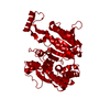
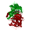
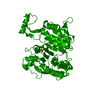
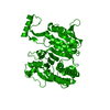
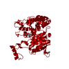
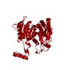
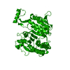
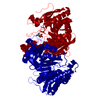
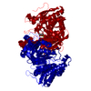
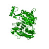
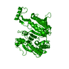
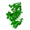
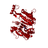
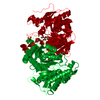
| Title | 3D structure of E. coli Isocitrate Dehydrogenase K100M mutant in complex with alpha-ketoglutarate, magnesium(II) and NADPH - The product complex | ||||||
 Components Components | ISOCITRATE DEHYDROGENASE [NADP] | ||||||
 Keywords Keywords | OXIDOREDUCTASE / OXIDATIVE BETA-DECARBOXYLATION | ||||||
| Function / homology |  Function and homology information Function and homology informationisocitrate dehydrogenase (NADP+) / isocitrate dehydrogenase (NADP+) activity / glyoxylate cycle / guanosine tetraphosphate binding / tricarboxylic acid cycle / electron transport chain / NAD binding / response to oxidative stress / magnesium ion binding / cytoplasm / cytosol Similarity search - Function | ||||||
| Biological species |  | ||||||
| Method |  X-RAY DIFFRACTION / SYNCHROTRON / MOLECULAR REPLACEMENT / Resolution: 2.687 Å X-RAY DIFFRACTION / SYNCHROTRON / MOLECULAR REPLACEMENT / Resolution: 2.687 Å | ||||||
 Authors Authors | Goncalves, S. / Miller, S.P. / Carrondo, M.A. / Dean, A.M. / Matias, P.M. | ||||||
 Citation Citation | Journal: Biochemistry / Year: 2012 Title: Induced Fit and the Catalytic Mechanism of Isocitrate Dehydrogenase. Authors: Goncalves, S. / Miller, S.P. / Carrondo, M.A. / Dean, A.M. / Matias, P.M. | ||||||
| History |
|
- Structure visualization
| Structure viewer | Molecule: MolmilJmol/JSmol |
|---|
- Downloads & links
-Download
| PDBx/mmCIF format | 4ajr.cif.gz | 100.7 KB | Display | PDBx/mmCIF format |
|---|---|---|---|---|
| PDB format | pdb4ajr.ent.gz | 74.8 KB | Display | PDB format |
| PDBx/mmJSON format | 4ajr.json.gz | Tree view | PDBx/mmJSON format | |
| Others |  Other downloads Other downloads |
-Validation report
| Arichive directory | https://data.pdbj.org/pub/pdb/validation_reports/aj/4ajrftp://data.pdbj.org/pub/pdb/validation_reports/aj/4ajr | HTTPS FTP |
|---|
-Related structure data
| Related structure data | 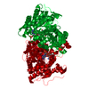 4aj3C 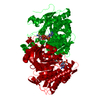 4ajaC  4ajbC 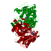 4ajcC  4ajsC 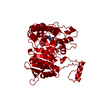 1ai2S S: Starting model for refinement C: citing same article ( |
|---|---|
| Similar structure data |


-Links
 PDBj
PDBj


- Assembly
| Deposited unit | 
| ||||||||
|---|---|---|---|---|---|---|---|---|---|
| 1 | 
| ||||||||
| Unit cell |
|
-Components
-Protein , 1 types, 1 molecules A
| #1: Protein | Mass: 45811.578 Da / Num. of mol.: 1 / Mutation: YES Source method: isolated from a genetically manipulated source Source: (gene. exp.) References: UniProt: P08200, isocitrate dehydrogenase (NADP+) |
|---|
-Non-polymers , 5 types, 131 molecules 


| #2: Chemical | ChemComp-NMN /  Mass: 335.227 Da / Num. of mol.: 1 / Source method: obtained synthetically / Formula: C11H16N2O8P Mass: 335.227 Da / Num. of mol.: 1 / Source method: obtained synthetically / Formula: C11H16N2O8P |
|---|---|
| #3: Chemical | ChemComp-NAP /  Mass: 743.405 Da / Num. of mol.: 1 / Source method: obtained synthetically / Formula: C21H28N7O17P3 Mass: 743.405 Da / Num. of mol.: 1 / Source method: obtained synthetically / Formula: C21H28N7O17P3 |
| #4: Chemical | ChemComp-AKG /  Mass: 146.098 Da / Num. of mol.: 1 / Source method: obtained synthetically / Formula: C5H6O5 Mass: 146.098 Da / Num. of mol.: 1 / Source method: obtained synthetically / Formula: C5H6O5 |
| #5: Chemical | ChemComp-MG /  Mass: 24.305 Da / Num. of mol.: 1 / Source method: obtained synthetically / Formula: Mg Mass: 24.305 Da / Num. of mol.: 1 / Source method: obtained synthetically / Formula: Mg |
| #6: Water | ChemComp-HOH / Mass: 18.015 Da / Num. of mol.: 127 / Source method: isolated from a natural source / Formula: H2O |
-Details
| Compound details | ENGINEERED| Nonpolymer details | NADP NICOTINAMIDE-ADENINE-DINUCLEOTIDE PHOSPHATE (NAP): THIS IS NADPH, THE REDUCED FORM OF NADP ...NADP NICOTINAMI | |
|---|
-Experimental details
-Experiment
| Experiment | Method: X-RAY DIFFRACTION / Number of used crystals: 1 |
|---|
- Sample preparation
| Crystal | Density Matthews: 4.5 Å3/Da / Density % sol: 72 % / Description: NONE |
|---|---|
| Crystal grow | Details: 1.85 M NH4SO4, 50 MM CITRIC ACID/NA2HPO4 BUFFER PH 5.2, 0.1 M NACL AND 0.2 M DTT |
-Data collection
| Diffraction | Mean temperature: 100 K |
|---|---|
| Diffraction source | Source: SYNCHROTRON / Site: ESRF  / Beamline: ID14-4 / Wavelength: 0.9535 / Beamline: ID14-4 / Wavelength: 0.9535 |
| Detector | Type: ADSC QUANTUM 315r / Detector: CCD / Details: TOROIDAL MIRROR |
| Radiation | Monochromator: CHANNEL-CUT ESRF MONOCHROMATOR / Protocol: SINGLE WAVELENGTH / Monochromatic (M) / Laue (L): M / Scattering type: x-ray |
| Radiation wavelength | Wavelength: 0.9535 Å / Relative weight: 1 |
| Reflection | Resolution: 2.69→60 Å / Num. obs: 23556 / % possible obs: 99.5 % / Observed criterion σ(I): -3 / Redundancy: 8.6 % / Biso Wilson estimate: 47.08 Å2 / Rmerge(I) obs: 0.12 / Net I/σ(I): 15.2 |
| Reflection shell | Resolution: 2.69→2.85 Å / Redundancy: 8.3 % / Rmerge(I) obs: 0.84 / Mean I/σ(I) obs: 2.8 / % possible all: 97.5 |
- Processing
| Software |
| ||||||||||||||||||||||||||||||||||||||||||||||||||||||||||||||||||||||
|---|---|---|---|---|---|---|---|---|---|---|---|---|---|---|---|---|---|---|---|---|---|---|---|---|---|---|---|---|---|---|---|---|---|---|---|---|---|---|---|---|---|---|---|---|---|---|---|---|---|---|---|---|---|---|---|---|---|---|---|---|---|---|---|---|---|---|---|---|---|---|---|
| Refinement | Method to determine structure: MOLECULAR REPLACEMENT Starting model: PDB ENTRY 1AI2 Resolution: 2.687→52.894 Å / SU ML: 0.66 / σ(F): 1.99 / Phase error: 18.02 / Stereochemistry target values: ML
| ||||||||||||||||||||||||||||||||||||||||||||||||||||||||||||||||||||||
| Solvent computation | Shrinkage radii: 0.83 Å / VDW probe radii: 1.1 Å / Solvent model: FLAT BULK SOLVENT MODEL / Bsol: 27.034 Å2 / ksol: 0.396 e/Å3 | ||||||||||||||||||||||||||||||||||||||||||||||||||||||||||||||||||||||
| Displacement parameters |
| ||||||||||||||||||||||||||||||||||||||||||||||||||||||||||||||||||||||
| Refinement step | Cycle: LAST / Resolution: 2.687→52.894 Å
| ||||||||||||||||||||||||||||||||||||||||||||||||||||||||||||||||||||||
| Refine LS restraints |
| ||||||||||||||||||||||||||||||||||||||||||||||||||||||||||||||||||||||
| LS refinement shell |
|
