 Movie
Movie Controller

[English] 日本語
 Yorodumi
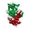
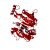
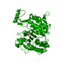
- PDB-1iso: ISOCITRATE DEHYDROGENASE: STRUCTURE OF AN ENGINEERED NADP+--> NAD... -
+ Open data

- Basic information
| Entry | Database: PDB / ID: 1iso | ||||||
|---|---|---|---|---|---|---|---|
| Title | ISOCITRATE DEHYDROGENASE: STRUCTURE OF AN ENGINEERED NADP+--> NAD+ SPECIFICITY-REVERSAL MUTANT | ||||||
 Components Components | ISOCITRATE DEHYDROGENASE | ||||||
 Keywords Keywords | OXIDOREDUCTASE / NADP / PHOSPHORYLATION / GLYOXYLATE BYPASS | ||||||
| Function / homology |  Function and homology information Function and homology informationisocitrate dehydrogenase (NADP+) / isocitrate dehydrogenase (NADP+) activity / glyoxylate cycle / guanosine tetraphosphate binding / tricarboxylic acid cycle / electron transport chain / NAD binding / response to oxidative stress / magnesium ion binding / cytosol / cytoplasm Similarity search - Function | ||||||
| Biological species |  | ||||||
| Method |  X-RAY DIFFRACTION / MOLECULAR REPLACEMENT / Resolution: 1.9 Å X-RAY DIFFRACTION / MOLECULAR REPLACEMENT / Resolution: 1.9 Å | ||||||
 Authors Authors | Hurley, J.H. | ||||||
 Citation Citation | Journal: Biochemistry / Year: 1996 Title: Determinants of cofactor specificity in isocitrate dehydrogenase: structure of an engineered NADP+ --> NAD+ specificity-reversal mutant. Authors: Hurley, J.H. / Chen, R. / Dean, A.M. | ||||||
| History |
|
- Structure visualization
| Structure viewer | Molecule: MolmilJmol/JSmol |
|---|
- Downloads & links
-Download
| PDBx/mmCIF format | 1iso.cif.gz | 102 KB | Display | PDBx/mmCIF format |
|---|---|---|---|---|
| PDB format | pdb1iso.ent.gz | 77.1 KB | Display | PDB format |
| PDBx/mmJSON format | 1iso.json.gz | Tree view | PDBx/mmJSON format | |
| Others |  Other downloads Other downloads |
-Validation report
| Arichive directory | https://data.pdbj.org/pub/pdb/validation_reports/is/1isoftp://data.pdbj.org/pub/pdb/validation_reports/is/1iso | HTTPS FTP |
|---|
-Related structure data
| Related structure data | 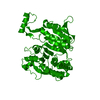 3icdS S: Starting model for refinement |
|---|---|
| Similar structure data |


-Links
 PDBj
PDBj

- Assembly
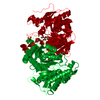
| Deposited unit | 
| ||||||||
|---|---|---|---|---|---|---|---|---|---|
| 1 | 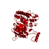
| ||||||||
| Unit cell |
|
-Components
| #1: Protein | Mass: 45701.379 Da / Num. of mol.: 1 / Mutation: C201M, C332Y, K344D, Y345I, V351A, Y391K, R395S Source method: isolated from a genetically manipulated source Source: (gene. exp.) References: UniProt: P08200, isocitrate dehydrogenase (NADP+) | ||||||
|---|---|---|---|---|---|---|---|
| #2: Chemical |   Mass: 96.063 Da / Num. of mol.: 3 / Source method: obtained synthetically / Formula: SO4 Mass: 96.063 Da / Num. of mol.: 3 / Source method: obtained synthetically / Formula: SO4#3: Chemical | ChemComp-NAD / |   Mass: 663.425 Da / Num. of mol.: 1 / Source method: obtained synthetically / Formula: C21H27N7O14P2 / Comment: NAD*YM Mass: 663.425 Da / Num. of mol.: 1 / Source method: obtained synthetically / Formula: C21H27N7O14P2 / Comment: NAD*YM#4: Water | ChemComp-HOH / |  Mass: 18.015 Da / Num. of mol.: 378 / Source method: isolated from a natural source / Formula: H2O Mass: 18.015 Da / Num. of mol.: 378 / Source method: isolated from a natural source / Formula: H2ONonpolymer details | ONLY AMP MOIETY OF THE NAD IS WELL-ORDERED. | |
-Experimental details
-Experiment
| Experiment | Method: X-RAY DIFFRACTION / Number of used crystals: 2 |
|---|
- Sample preparation
| Crystal | Density Matthews: 4.32 Å3/Da / Density % sol: 71.51 % | ||||||||||||||||||||||||||||||||||||||||||
|---|---|---|---|---|---|---|---|---|---|---|---|---|---|---|---|---|---|---|---|---|---|---|---|---|---|---|---|---|---|---|---|---|---|---|---|---|---|---|---|---|---|---|---|
| Crystal grow | pH: 5.8 / Details: pH 5.8 | ||||||||||||||||||||||||||||||||||||||||||
| Crystal grow | *PLUS pH: 5.4 / Method: unknown | ||||||||||||||||||||||||||||||||||||||||||
| Components of the solutions | *PLUS
|
-Data collection
| Diffraction | Mean temperature: 93 K |
|---|---|
| Diffraction source | Source: ROTATING ANODE / Type: RIGAKU RUH2R / Wavelength: 1.5418 |
| Detector | Type: RIGAKU RAXIS IIC / Detector: IMAGE PLATE / Details: MIRRORS |
| Radiation | Monochromatic (M) / Laue (L): M / Scattering type: x-ray |
| Radiation wavelength | Wavelength: 1.5418 Å / Relative weight: 1 |
| Reflection | Resolution: 1.9→100 Å / Num. obs: 63350 / % possible obs: 99.1 % / Observed criterion σ(I): -1 / Rmerge(I) obs: 0.055 / Net I/σ(I): 22.6 |
| Reflection shell | Resolution: 1.09→1.97 Å / Rmerge(I) obs: 0.27 / Mean I/σ(I) obs: 4.5 / % possible all: 97 |
| Reflection shell | *PLUS % possible obs: 97 % / Num. unique obs: 6069 |
- Processing
| Software |
| ||||||||||||||||||||||||||||||||||||||||||||||||||||||||||||
|---|---|---|---|---|---|---|---|---|---|---|---|---|---|---|---|---|---|---|---|---|---|---|---|---|---|---|---|---|---|---|---|---|---|---|---|---|---|---|---|---|---|---|---|---|---|---|---|---|---|---|---|---|---|---|---|---|---|---|---|---|---|
| Refinement | Method to determine structure: MOLECULAR REPLACEMENT Starting model: 3ICD Resolution: 1.9→6 Å / σ(F): 0
| ||||||||||||||||||||||||||||||||||||||||||||||||||||||||||||
| Displacement parameters | Biso mean: 16.5 Å2 | ||||||||||||||||||||||||||||||||||||||||||||||||||||||||||||
| Refinement step | Cycle: LAST / Resolution: 1.9→6 Å
| ||||||||||||||||||||||||||||||||||||||||||||||||||||||||||||
| Refine LS restraints |
| ||||||||||||||||||||||||||||||||||||||||||||||||||||||||||||
| Xplor file |
| ||||||||||||||||||||||||||||||||||||||||||||||||||||||||||||
| Software | *PLUS Name: X-PLOR / Classification: refinement | ||||||||||||||||||||||||||||||||||||||||||||||||||||||||||||
| Refinement | *PLUS | ||||||||||||||||||||||||||||||||||||||||||||||||||||||||||||
| Solvent computation | *PLUS | ||||||||||||||||||||||||||||||||||||||||||||||||||||||||||||
| Displacement parameters | *PLUS | ||||||||||||||||||||||||||||||||||||||||||||||||||||||||||||
| Refine LS restraints | *PLUS
|
