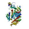
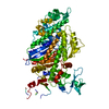
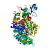
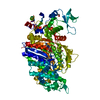
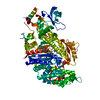
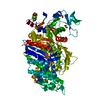
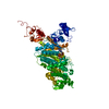
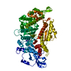
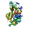
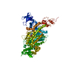
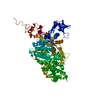
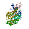
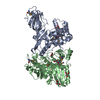
Resolution: 2.5→41.914 Å / SU ML: 0.35 / σ(F): 0 / Phase error: 25.63 / Stereochemistry target values: ML Details: LEVER ARM RESIDUES(FROM 700 TO 748) ARE HIGHLY MOBILE.
Rfactor
Num. reflection
% reflection
Rfree
0.2536
1721
5 %
Rwork
0.2138
-
-
obs
0.2158
34133
98.83 %
Solvent computation
Shrinkage radii: 0.9 Å / VDW probe radii: 1.11 Å / Solvent model: FLAT BULK SOLVENT MODEL / Bsol: 63.103 Å2 / ksol: 0.35 e/Å3
Displacement parameters
Baniso -1
Baniso -2
Baniso -3
1-
3.5819 Å2
0 Å2
0 Å2
2-
-
6.4747 Å2
0 Å2
3-
-
-
-10.0566 Å2
Refinement step
Cycle: LAST / Resolution: 2.5→41.914 Å
Protein
Nucleic acid
Ligand
Solvent
Total
Num. atoms
5963
0
89
439
6491
Refine LS restraints
Refine-ID
Type
Dev ideal
Number
X-RAY DIFFRACTION
f_bond_d
0.012
6172
X-RAY DIFFRACTION
f_angle_d
1.554
8340
X-RAY DIFFRACTION
f_dihedral_angle_d
15.92
2334
X-RAY DIFFRACTION
f_chiral_restr
0.103
902
X-RAY DIFFRACTION
f_plane_restr
0.009
1076
LS refinement shell
Resolution (Å)
Rfactor Rfree
Num. reflection Rfree
Rfactor Rwork
Num. reflection Rwork
Refine-ID
% reflection obs (%)
2.5-2.5736
0.3449
132
0.2711
2701
X-RAY DIFFRACTION
100
2.5736-2.6566
0.2573
117
0.2519
2711
X-RAY DIFFRACTION
99
2.6566-2.7515
0.3001
161
0.2479
2675
X-RAY DIFFRACTION
99
2.7515-2.8617
0.3602
131
0.2574
2658
X-RAY DIFFRACTION
99
2.8617-2.9919
0.3409
153
0.2542
2673
X-RAY DIFFRACTION
100
2.9919-3.1496
0.294
148
0.2264
2686
X-RAY DIFFRACTION
98
3.1496-3.3468
0.2385
140
0.2269
2641
X-RAY DIFFRACTION
98
3.3468-3.6051
0.2356
140
0.2002
2667
X-RAY DIFFRACTION
98
3.6051-3.9677
0.2255
150
0.1926
2668
X-RAY DIFFRACTION
98
3.9677-4.5412
0.2137
139
0.1771
2704
X-RAY DIFFRACTION
98
4.5412-5.7191
0.2327
168
0.1951
2745
X-RAY DIFFRACTION
99
5.7191-41.9199
0.2403
142
0.2151
2883
X-RAY DIFFRACTION
100
+
About Yorodumi
-
News
-
Feb 9, 2022. New format data for meta-information of EMDB entries
New format data for meta-information of EMDB entries
Version 3 of the EMDB header file is now the official format.
The previous official version 1.9 will be removed from the archive.
In the structure databanks used in Yorodumi, some data are registered as the other names, "COVID-19 virus" and "2019-nCoV". Here are the details of the virus and the list of structure data.
Jan 31, 2019. EMDB accession codes are about to change! (news from PDBe EMDB page)
EMDB accession codes are about to change! (news from PDBe EMDB page)
The allocation of 4 digits for EMDB accession codes will soon come to an end. Whilst these codes will remain in use, new EMDB accession codes will include an additional digit and will expand incrementally as the available range of codes is exhausted. The current 4-digit format prefixed with “EMD-” (i.e. EMD-XXXX) will advance to a 5-digit format (i.e. EMD-XXXXX), and so on. It is currently estimated that the 4-digit codes will be depleted around Spring 2019, at which point the 5-digit format will come into force.
The EM Navigator/Yorodumi systems omit the EMD- prefix.
Related info.:Q: What is EMD? / ID/Accession-code notation in Yorodumi/EM Navigator
Yorodumi is a browser for structure data from EMDB, PDB, SASBDB, etc.
This page is also the successor to EM Navigator detail page, and also detail information page/front-end page for Omokage search.
The word "yorodu" (or yorozu) is an old Japanese word meaning "ten thousand". "mi" (miru) is to see.
Related info.:EMDB / PDB / SASBDB / Comparison of 3 databanks / Yorodumi Search / Aug 31, 2016. New EM Navigator & Yorodumi / Yorodumi Papers / Jmol/JSmol / Function and homology information / Changes in new EM Navigator and Yorodumi
 Movie
Movie Controller

 Yorodumi
 Open data
 Basic information
 Components
 Keywords
 Function and homology information
 X-RAY DIFFRACTION /
  Authors
 Citation
 Structure visualization
 Downloads & links
 Other downloads
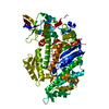


 PDBj
PDBj


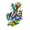
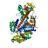
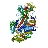
 Assembly
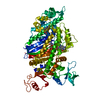
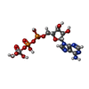


 Mass: 544.156 Da / Num. of mol.: 1 / Source method: obtained synthetically / Formula: C10H17N5O14P2V / Comment: energy-carrying molecule analogue*YM
 Mass: 24.305 Da / Num. of mol.: 1 / Source method: obtained synthetically / Formula: Mg
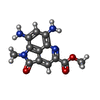
 Mass: 272.259 Da / Num. of mol.: 2 / Source method: obtained synthetically / Formula: C13H12N4O3
 Mass: 62.068 Da / Num. of mol.: 4 / Source method: obtained synthetically / Formula: C2H6O2
 Sample preparation
 Processing