 Movie
Movie Controller

[English] 日本語
 Yorodumi
- PDB-1w9j: Myosin II Dictyostelium discoideum motor domain S456Y bound with ... -
+ Open data

- Basic information
| Entry | Database: PDB / ID: 1w9j | ||||||
|---|---|---|---|---|---|---|---|
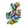
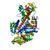
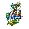
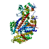
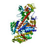
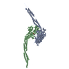
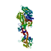
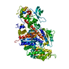
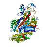
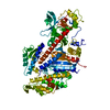
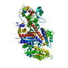
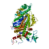
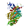
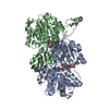
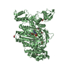
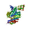
| Title | Myosin II Dictyostelium discoideum motor domain S456Y bound with MgADP-AlF4 | ||||||
 Components Components | MYOSIN II HEAVY CHAIN | ||||||
 Keywords Keywords | MYOSIN / MOLECULAR MOTOR / ATPASE / MOTOR DOMAIN / MUTANT / MUSCLE CONTRACTION | ||||||
| Function / homology |  Function and homology information Function and homology informationuropod retraction / cytoplasmic actin-based contraction involved in forward cell motility / phagocytic cup base / pathogen-containing vacuole / response to differentiation-inducing factor 1 / equatorial cell cortex / contractile actin filament bundle assembly / pseudopodium retraction / cell trailing edge / contractile vacuole organization ...uropod retraction / cytoplasmic actin-based contraction involved in forward cell motility / phagocytic cup base / pathogen-containing vacuole / response to differentiation-inducing factor 1 / equatorial cell cortex / contractile actin filament bundle assembly / pseudopodium retraction / cell trailing edge / contractile vacuole organization / myosin filament assembly / aggregation involved in sorocarp development / culmination involved in sorocarp development / adenyl nucleotide binding / RHO GTPases activate PAKs / calcium-dependent ATPase activity / actomyosin contractile ring / hypotonic response / uropod / apical cortex / negative regulation of actin filament polymerization / actin-myosin filament sliding / bleb assembly / detection of mechanical stimulus / substrate-dependent cell migration, cell extension / actomyosin / filopodium assembly / early phagosome / myosin filament / cortical actin cytoskeleton organization / myosin II complex / cortical actin cytoskeleton / microfilament motor activity / cleavage furrow / pseudopodium / mitotic cytokinesis / cytoskeletal motor activity / response to cAMP / response to mechanical stimulus / 14-3-3 protein binding / cell motility / response to hydrogen peroxide / chemotaxis / actin filament binding / intracellular protein localization / regulation of cell shape / extracellular matrix / cytoplasmic vesicle / cell cortex / cytoskeleton / calmodulin binding / ATP binding / identical protein binding / cytosol / cytoplasm Similarity search - Function | ||||||
| Biological species |  | ||||||
| Method |  X-RAY DIFFRACTION / SYNCHROTRON / MOLECULAR REPLACEMENT / Resolution: 2 Å X-RAY DIFFRACTION / SYNCHROTRON / MOLECULAR REPLACEMENT / Resolution: 2 Å | ||||||
 Authors Authors | Morris, C.A. / Coureux, P.-D. / Wells, A.L. / Houdusse, A. / Sweeney, H.L. | ||||||
 Citation Citation | Journal: To be Published Title: Structure-Function Analysis of Myosin II Backdoor Mutants Authors: Morris, C.A. / Coureux, P.-D. / Wells, A.L. / Houdusse, A. / Sweeney, H.L. | ||||||
| History |
|
- Structure visualization
| Structure viewer | Molecule: MolmilJmol/JSmol |
|---|
- Downloads & links
-Download
| PDBx/mmCIF format | 1w9j.cif.gz | 168.8 KB | Display | PDBx/mmCIF format |
|---|---|---|---|---|
| PDB format | pdb1w9j.ent.gz | 129.9 KB | Display | PDB format |
| PDBx/mmJSON format | 1w9j.json.gz | Tree view | PDBx/mmJSON format | |
| Others |  Other downloads Other downloads |
-Validation report
| Arichive directory | https://data.pdbj.org/pub/pdb/validation_reports/w9/1w9jftp://data.pdbj.org/pub/pdb/validation_reports/w9/1w9j | HTTPS FTP |
|---|
-Related structure data
| Related structure data |  1w9iC  1w9kC  1w9lC  1mndS S: Starting model for refinement C: citing same article ( |
|---|---|
| Similar structure data |


-Links
 PDBj
PDBj


- Assembly
| Deposited unit | 
| ||||||||
|---|---|---|---|---|---|---|---|---|---|
| 1 |
| ||||||||
| Unit cell |
|
-Components
-Protein , 1 types, 1 molecules A
| #1: Protein | Mass: 87897.344 Da / Num. of mol.: 1 / Fragment: MOTOR DOMAIN, RESIDUES 1-755 / Mutation: YES Source method: isolated from a genetically manipulated source Details: MGADP-BEFX / Source: (gene. exp.)   SPODOPTERA FRUGIPERDA (fall armyworm) / References: UniProt: P08799, EC: 3.6.4.1 SPODOPTERA FRUGIPERDA (fall armyworm) / References: UniProt: P08799, EC: 3.6.4.1 |
|---|
-Non-polymers , 5 types, 397 molecules 


| #2: Chemical | ChemComp-MG /  Mass: 24.305 Da / Num. of mol.: 1 / Source method: obtained synthetically / Formula: Mg Mass: 24.305 Da / Num. of mol.: 1 / Source method: obtained synthetically / Formula: Mg | ||
|---|---|---|---|
| #3: Chemical | ChemComp-ADP /  Mass: 427.201 Da / Num. of mol.: 1 / Source method: obtained synthetically / Formula: C10H15N5O10P2 / Comment: ADP, energy-carrying molecule*YM Mass: 427.201 Da / Num. of mol.: 1 / Source method: obtained synthetically / Formula: C10H15N5O10P2 / Comment: ADP, energy-carrying molecule*YM | ||
| #4: Chemical | ChemComp-ALF /  Mass: 102.975 Da / Num. of mol.: 1 / Source method: obtained synthetically / Formula: AlF4 Mass: 102.975 Da / Num. of mol.: 1 / Source method: obtained synthetically / Formula: AlF4 | ||
| #5: Chemical | ChemComp-EDO /  Mass: 62.068 Da / Num. of mol.: 5 / Source method: obtained synthetically / Formula: C2H6O2 Mass: 62.068 Da / Num. of mol.: 5 / Source method: obtained synthetically / Formula: C2H6O2#6: Water | ChemComp-HOH / | Mass: 18.015 Da / Num. of mol.: 389 / Source method: isolated from a natural source / Formula: H2O |
-Details
| Compound details | ENGINEERED RESIDUE ASP 2 ASN, CHAIN A ENGINEERED RESIDUE SER 456 TYR, CHAIN A MYOSIN IS A PROTEIN ...ENGINEERED |
|---|
-Experimental details
-Experiment
| Experiment | Method: X-RAY DIFFRACTION / Number of used crystals: 1 |
|---|
- Sample preparation
| Crystal | Density Matthews: 3.1 Å3/Da / Density % sol: 59.4 % |
|---|---|
| Crystal grow | pH: 7 Details: 12% PEG 8000, 50 MM HEPES PH 7, 100 MM NACL, 2 MM DTT, 2 MM NAN3 |
-Data collection
| Diffraction | Mean temperature: 100 K |
|---|---|
| Diffraction source | Source: SYNCHROTRON / Site: ESRF  / Beamline: BM30A / Wavelength: 0.9793 / Beamline: BM30A / Wavelength: 0.9793 |
| Detector | Type: MARRESEARCH / Detector: IMAGE PLATE / Date: Jun 24, 2000 |
| Radiation | Protocol: SINGLE WAVELENGTH / Monochromatic (M) / Laue (L): M / Scattering type: x-ray |
| Radiation wavelength | Wavelength: 0.9793 Å / Relative weight: 1 |
| Reflection | Resolution: 2→76.7 Å / Num. obs: 63165 / % possible obs: 89.4 % / Observed criterion σ(I): 3 / Redundancy: 5.4 % / Rmerge(I) obs: 0.07 / Net I/σ(I): 20.3 |
| Reflection shell | Resolution: 1.98→2.03 Å / Redundancy: 3.7 % / Rmerge(I) obs: 0.21 / Mean I/σ(I) obs: 2.9 / % possible all: 44.4 |
- Processing
| Software |
| ||||||||||||||||||||||||||||||||||||||||||||||||||||||||||||||||||||||||||||||||||||||||||||||||||||||||||||||||||||||||||||||||||||||||||||||||||||||||||||||||||||||||||||||||||||||
|---|---|---|---|---|---|---|---|---|---|---|---|---|---|---|---|---|---|---|---|---|---|---|---|---|---|---|---|---|---|---|---|---|---|---|---|---|---|---|---|---|---|---|---|---|---|---|---|---|---|---|---|---|---|---|---|---|---|---|---|---|---|---|---|---|---|---|---|---|---|---|---|---|---|---|---|---|---|---|---|---|---|---|---|---|---|---|---|---|---|---|---|---|---|---|---|---|---|---|---|---|---|---|---|---|---|---|---|---|---|---|---|---|---|---|---|---|---|---|---|---|---|---|---|---|---|---|---|---|---|---|---|---|---|---|---|---|---|---|---|---|---|---|---|---|---|---|---|---|---|---|---|---|---|---|---|---|---|---|---|---|---|---|---|---|---|---|---|---|---|---|---|---|---|---|---|---|---|---|---|---|---|---|---|
| Refinement | Method to determine structure: MOLECULAR REPLACEMENT Starting model: PDB ENTRY 1MND Resolution: 2→76.7 Å / Cor.coef. Fo:Fc: 0.951 / Cor.coef. Fo:Fc free: 0.935 / SU B: 4.04 / SU ML: 0.111 / Cross valid method: THROUGHOUT / ESU R: 0.172 / ESU R Free: 0.153 / Stereochemistry target values: MAXIMUM LIKELIHOOD / Details: HYDROGENS HAVE BEEN ADDED IN THE RIDING POSITIONS.
| ||||||||||||||||||||||||||||||||||||||||||||||||||||||||||||||||||||||||||||||||||||||||||||||||||||||||||||||||||||||||||||||||||||||||||||||||||||||||||||||||||||||||||||||||||||||
| Solvent computation | Ion probe radii: 0.8 Å / Shrinkage radii: 0.8 Å / VDW probe radii: 1.4 Å / Solvent model: BABINET MODEL WITH MASK | ||||||||||||||||||||||||||||||||||||||||||||||||||||||||||||||||||||||||||||||||||||||||||||||||||||||||||||||||||||||||||||||||||||||||||||||||||||||||||||||||||||||||||||||||||||||
| Displacement parameters | Biso mean: 37.25 Å2
| ||||||||||||||||||||||||||||||||||||||||||||||||||||||||||||||||||||||||||||||||||||||||||||||||||||||||||||||||||||||||||||||||||||||||||||||||||||||||||||||||||||||||||||||||||||||
| Refinement step | Cycle: LAST / Resolution: 2→76.7 Å
| ||||||||||||||||||||||||||||||||||||||||||||||||||||||||||||||||||||||||||||||||||||||||||||||||||||||||||||||||||||||||||||||||||||||||||||||||||||||||||||||||||||||||||||||||||||||
| Refine LS restraints |
|