[English] 日本語
Yorodumi
- PDB-5i0h: Crystal structure of myosin X motor domain in pre-powerstroke state -

+
Open data


ID or keywords:

Loading...

-
Basic information

Entry
Database: PDB / ID: 5i0h
TitleCrystal structure of myosin X motor domain in pre-powerstroke state
ComponentsUnconventional myosin-X
KeywordsMOTOR PROTEIN / myosin / motor domain / molecular motor / pre-powerstroke state / motility
Function / homology
Function and homology information


plus-end directed microfilament motor activity / Netrin-1 signaling / positive regulation of cell-cell adhesion / filopodium tip / regulation of filopodium assembly / cytoskeleton-dependent intracellular transport / filopodium membrane / myosin complex / spectrin binding / microfilament motor activity ...plus-end directed microfilament motor activity / Netrin-1 signaling / positive regulation of cell-cell adhesion / filopodium tip / regulation of filopodium assembly / cytoskeleton-dependent intracellular transport / filopodium membrane / myosin complex / spectrin binding / microfilament motor activity / phosphatidylinositol-3,4,5-trisphosphate binding / ruffle / filopodium / FCGR3A-mediated phagocytosis / Regulation of actin dynamics for phagocytic cup formation / actin filament binding / lamellipodium / cell cortex / regulation of cell shape / calmodulin binding / neuron projection / neuronal cell body / nucleolus / signal transduction / ATP binding / plasma membrane / cytosol
Similarity search - Function
Unconventional myosin-X, coiled coil domain / Class X myosin, motor domain / Myosin X, N-terminal SH3 domain / Myosin X, FERM domain C-lobe / Unconventional myosin-X coiled coil domain / Myosin X N-terminal SH3 domain / MyTH4 domain / MyTH4 domain superfamily / MyTH4 domain / MyTH4 domain profile. ...Unconventional myosin-X, coiled coil domain / Class X myosin, motor domain / Myosin X, N-terminal SH3 domain / Myosin X, FERM domain C-lobe / Unconventional myosin-X coiled coil domain / Myosin X N-terminal SH3 domain / MyTH4 domain / MyTH4 domain superfamily / MyTH4 domain / MyTH4 domain profile. / Domain in Myosin and Kinesin Tails / Ras association (RalGDS/AF-6) domain / Ras-associating (RA) domain / IQ calmodulin-binding motif / FERM central domain / FERM/acyl-CoA-binding protein superfamily / Short calmodulin-binding motif containing conserved Ile and Gln residues. / Myosin head, motor domain / Myosin head (motor domain) / Myosin motor domain profile. / Myosin. Large ATPases. / IQ motif profile. / IQ motif, EF-hand binding site / FERM central domain / FERM superfamily, second domain / FERM domain / FERM domain profile. / Band 4.1 domain / Band 4.1 homologues / Kinesin motor domain superfamily / PH domain / PH domain profile. / Pleckstrin homology domain. / Pleckstrin homology domain / PH-like domain superfamily / P-loop containing nucleoside triphosphate hydrolase
Similarity search - Domain/homology
ADENOSINE-5'-DIPHOSPHATE / BERYLLIUM TRIFLUORIDE ION / Unconventional myosin-X
Similarity search - Component
Biological speciesHomo sapiens (human)
MethodX-RAY DIFFRACTION / SYNCHROTRON / MOLECULAR REPLACEMENT / Resolution: 1.8 Å
AuthorsIsabet, T. / Blanc, F. / Sweeney, H.L. / Houdusse, A.
Funding support France, United States, 6items
OrganizationGrant numberCountry
French National Research AgencyANR blanche BLAN10 France
French National Research AgencyANR-13-BSV8-0019-01 France
Ligue contre le cancer France
ARC France
National Institutes of HealthDC009100 United States
National Institutes of HealthHL110869 United States
Citation
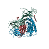
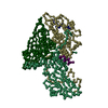
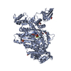
Journal: Nat Commun / Year: 2016
Title: The myosin X motor is optimized for movement on actin bundles.
Authors: Virginie Ropars / Zhaohui Yang / Tatiana Isabet / Florian Blanc / Kaifeng Zhou / Tianming Lin / Xiaoyan Liu / Pascale Hissier / Frédéric Samazan / Béatrice Amigues / Eric D Yang / Hyokeun ...Authors: Virginie Ropars / Zhaohui Yang / Tatiana Isabet / Florian Blanc / Kaifeng Zhou / Tianming Lin / Xiaoyan Liu / Pascale Hissier / Frédéric Samazan / Béatrice Amigues / Eric D Yang / Hyokeun Park / Olena Pylypenko / Marco Cecchini / Charles V Sindelar / H Lee Sweeney / Anne Houdusse /
Abstract: Myosin X has features not found in other myosins. Its structure must underlie its unique ability to generate filopodia, which are essential for neuritogenesis, wound healing, cancer metastasis and ...Myosin X has features not found in other myosins. Its structure must underlie its unique ability to generate filopodia, which are essential for neuritogenesis, wound healing, cancer metastasis and some pathogenic infections. By determining high-resolution structures of key components of this motor, and characterizing the in vitro behaviour of the native dimer, we identify the features that explain the myosin X dimer behaviour. Single-molecule studies demonstrate that a native myosin X dimer moves on actin bundles with higher velocities and takes larger steps than on single actin filaments. The largest steps on actin bundles are larger than previously reported for artificially dimerized myosin X constructs or any other myosin. Our model and kinetic data explain why these large steps and high velocities can only occur on bundled filaments. Thus, myosin X functions as an antiparallel dimer in cells with a unique geometry optimized for movement on actin bundles.
History
DepositionFeb 4, 2016Deposition site: RCSB / Processing site: PDBE
Revision 1.0Sep 7, 2016Provider: repository / Type: Initial release
Revision 1.1Sep 14, 2016Group: Database references
Revision 1.2Aug 30, 2017Group: Author supporting evidence / Category: pdbx_audit_support / Item: _pdbx_audit_support.funding_organization
Revision 1.3Oct 16, 2019Group: Data collection / Category: reflns_shell
Revision 1.4May 8, 2024Group: Data collection / Database references / Derived calculations
Category: chem_comp_atom / chem_comp_bond ...chem_comp_atom / chem_comp_bond / database_2 / pdbx_struct_conn_angle / struct_conn
Item: _database_2.pdbx_DOI / _database_2.pdbx_database_accession ..._database_2.pdbx_DOI / _database_2.pdbx_database_accession / _pdbx_struct_conn_angle.ptnr1_auth_asym_id / _pdbx_struct_conn_angle.ptnr1_auth_comp_id / _pdbx_struct_conn_angle.ptnr1_auth_seq_id / _pdbx_struct_conn_angle.ptnr1_label_asym_id / _pdbx_struct_conn_angle.ptnr1_label_atom_id / _pdbx_struct_conn_angle.ptnr1_label_comp_id / _pdbx_struct_conn_angle.ptnr1_label_seq_id / _pdbx_struct_conn_angle.ptnr2_auth_asym_id / _pdbx_struct_conn_angle.ptnr2_auth_comp_id / _pdbx_struct_conn_angle.ptnr2_auth_seq_id / _pdbx_struct_conn_angle.ptnr2_label_asym_id / _pdbx_struct_conn_angle.ptnr2_label_atom_id / _pdbx_struct_conn_angle.ptnr2_label_comp_id / _pdbx_struct_conn_angle.ptnr3_auth_asym_id / _pdbx_struct_conn_angle.ptnr3_auth_comp_id / _pdbx_struct_conn_angle.ptnr3_auth_seq_id / _pdbx_struct_conn_angle.ptnr3_label_asym_id / _pdbx_struct_conn_angle.ptnr3_label_atom_id / _pdbx_struct_conn_angle.ptnr3_label_comp_id / _pdbx_struct_conn_angle.ptnr3_label_seq_id / _pdbx_struct_conn_angle.value / _struct_conn.pdbx_dist_value / _struct_conn.ptnr1_auth_asym_id / _struct_conn.ptnr1_auth_comp_id / _struct_conn.ptnr1_auth_seq_id / _struct_conn.ptnr1_label_asym_id / _struct_conn.ptnr1_label_atom_id / _struct_conn.ptnr1_label_comp_id / _struct_conn.ptnr1_label_seq_id / _struct_conn.ptnr2_auth_asym_id / _struct_conn.ptnr2_auth_comp_id / _struct_conn.ptnr2_auth_seq_id / _struct_conn.ptnr2_label_asym_id / _struct_conn.ptnr2_label_atom_id / _struct_conn.ptnr2_label_comp_id

-
Structure visualization

Structure viewerMolecule:
MolmilJmol/JSmol

Downloads & links

-
Assembly

Deposited unit
A: Unconventional myosin-X
B: Unconventional myosin-X
hetero molecules


Theoretical massNumber of molelcules
Total (without water)173,87031
Polymers171,2382
Non-polymers2,63329
Water22,6631258
1
A: Unconventional myosin-X
hetero molecules


Theoretical massNumber of molelcules
Total (without water)86,72913
Polymers85,6191
Non-polymers1,11012
Water181
TypeNameSymmetry operationNumber
identity operation1_555x,y,z1
2
B: Unconventional myosin-X
hetero molecules


Theoretical massNumber of molelcules
Total (without water)87,14118
Polymers85,6191
Non-polymers1,52217
Water181
TypeNameSymmetry operationNumber
identity operation1_555x,y,z1
Unit cell
Length a, b, c (Å)76.480, 78.300, 78.670
Angle α, β, γ (deg.)75.69, 86.38, 76.12
Int Tables number1
Space group name H-MP1

-
Components

-
Protein , 1 types, 2 molecules AB

#1: Protein Unconventional myosin-X / Unconventional myosin-10


Mass: 85618.781 Da / Num. of mol.: 2
Source method: isolated from a genetically manipulated source
Source: (gene. exp.) Homo sapiens (human) / Gene: MYO10, KIAA0799 / Production host: 
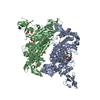
unidentified baculovirus / References: UniProt: Q9HD67

-
Non-polymers , 6 types, 1287 molecules

#2: Chemical ChemComp-ADP / ADENOSINE-5'-DIPHOSPHATE


Mass: 427.201 Da / Num. of mol.: 2 / Source method: obtained synthetically / Formula: C10H15N5O10P2 / Comment: ADP, energy-carrying molecule*YM
#3: Chemical ChemComp-MG / MAGNESIUM ION


Mass: 24.305 Da / Num. of mol.: 2 / Source method: obtained synthetically / Formula: Mg
#4: Chemical ChemComp-BEF / BERYLLIUM TRIFLUORIDE ION


Mass: 66.007 Da / Num. of mol.: 2 / Source method: obtained synthetically / Formula: BeF3
#5: Chemical
ChemComp-EDO / 1,2-ETHANEDIOL / ETHYLENE GLYCOL


Mass: 62.068 Da / Num. of mol.: 18 / Source method: obtained synthetically / Formula: C2H6O2
#6: Chemical
ChemComp-SO4 / SULFATE ION


Mass: 96.063 Da / Num. of mol.: 5 / Source method: obtained synthetically / Formula: SO4
#7: Water ChemComp-HOH / water


Mass: 18.015 Da / Num. of mol.: 1258 / Source method: isolated from a natural source / Formula: H2O

-
Experimental details

-
Experiment

ExperimentMethod: X-RAY DIFFRACTION / Number of used crystals: 1

-
Sample preparation

CrystalDensity Matthews: 2.59 Å3/Da / Density % sol: 52.47 %
Crystal growTemperature: 277 K / Method: vapor diffusion, hanging drop / pH: 7.5
Details: 10% PEG 8000, 50mM Tris pH 7.5, 1mM TCEP and 125mM Lithium sulfate

-
Data collection

DiffractionMean temperature: 100 K
Diffraction sourceSource: SYNCHROTRON / Site: SOLEIL / Beamline: PROXIMA 1 / Wavelength: 0.9798 Å
DetectorType: DECTRIS PILATUS 6M / Detector: PIXEL / Date: Feb 1, 2012
RadiationProtocol: SINGLE WAVELENGTH / Monochromatic (M) / Laue (L): M / Scattering type: x-ray
Radiation wavelengthWavelength: 0.9798 Å / Relative weight: 1
ReflectionResolution: 1.8→50 Å / Num. obs: 155150 / % possible obs: 97.4 % / Redundancy: 4.8 % / Biso Wilson estimate: 30.13 Å2 / Net I/σ(I): 15.96

-
Processing

Software
NameVersionClassification
BUSTER2.10.2refinement
XDSdata reduction
PHASERphasing
RefinementMethod to determine structure: MOLECULAR REPLACEMENT / Resolution: 1.8→26.35 Å / Cor.coef. Fo:Fc: 0.9616 / Cor.coef. Fo:Fc free: 0.9535 / SU R Cruickshank DPI: 0.108 / Cross valid method: THROUGHOUT / σ(F): 0 / SU R Blow DPI: 0.11 / SU Rfree Blow DPI: 0.101 / SU Rfree Cruickshank DPI: 0.101
RfactorNum. reflection% reflectionSelection details
Rfree0.1899 7758 5 %RANDOM
Rwork0.1654 ---
obs0.1666 155150 97.5 %-
Displacement parametersBiso mean: 37.11 Å2
Baniso -1Baniso -2Baniso -3
1-0.9805 Å2-3.7505 Å2-0.9214 Å2
2---1.4854 Å2-2.1684 Å2
3---0.5049 Å2
Refine analyzeLuzzati coordinate error obs: 0.204 Å
Refinement stepCycle: 1 / Resolution: 1.8→26.35 Å
ProteinNucleic acidLigandSolventTotal
Num. atoms11854 0 161 1258 13273
Refine LS restraints
Refine-IDTypeDev idealNumberRestraint functionWeight
X-RAY DIFFRACTIONt_bond_d0.0112260HARMONIC2
X-RAY DIFFRACTIONt_angle_deg116592HARMONIC2
X-RAY DIFFRACTIONt_dihedral_angle_d4350SINUSOIDAL2
X-RAY DIFFRACTIONt_incorr_chiral_ct
X-RAY DIFFRACTIONt_pseud_angle
X-RAY DIFFRACTIONt_trig_c_planes342HARMONIC2
X-RAY DIFFRACTIONt_gen_planes1814HARMONIC5
X-RAY DIFFRACTIONt_it12260HARMONIC20
X-RAY DIFFRACTIONt_nbd0SEMIHARMONIC5
X-RAY DIFFRACTIONt_omega_torsion3.32
X-RAY DIFFRACTIONt_other_torsion16.28
X-RAY DIFFRACTIONt_improper_torsion
X-RAY DIFFRACTIONt_chiral_improper_torsion1560SEMIHARMONIC5
X-RAY DIFFRACTIONt_sum_occupancies
X-RAY DIFFRACTIONt_utility_distance
X-RAY DIFFRACTIONt_utility_angle
X-RAY DIFFRACTIONt_utility_torsion
X-RAY DIFFRACTIONt_ideal_dist_contact15609SEMIHARMONIC4
LS refinement shellResolution: 1.8→1.85 Å / Total num. of bins used: 20
RfactorNum. reflection% reflection
Rfree0.2471 562 5.01 %
Rwork0.2017 10666 -
all0.2039 11228 -
obs--95.31 %
Refinement TLS params.

Method: refined / Refine-ID: X-RAY DIFFRACTION

IDL112)L122)L132)L222)L232)L332)S11 (Å °)S12 (Å °)S13 (Å °)S21 (Å °)S22 (Å °)S23 (Å °)S31 (Å °)S32 (Å °)S33 (Å °)T112)T122)T132)T222)T232)T332)Origin x (Å)Origin y (Å)Origin z (Å)
11.5997-0.3333-0.84361.41841.57996.42450.06890.22240.0342-0.16130.00470.1086-0.16810.1079-0.07360.06750.0412-0.03210.1737-0.0699-0.02044.7621-10.9192-26.3886
20.6246-0.24940.02340.6912-0.36690.6265-0.02890.008-0.12090.09670.07460.1302-0.0234-0.0191-0.0457-0.1258-0.00990.0108-0.1474-0.027-0.1347-2.3171-11.517813.9034
39.43542.08082.34918.96863.58229.03990.0927-1.7576-0.21860.9126-0.47430.82820.4149-1.43420.38160.06450.07020.23520.44790.12620.1326-54.5995-10.245960.481
40.5068-0.1865-0.08520.7272-0.3960.6838-0.0529-0.1124-0.02590.08530.00820.0026-0.03290.03110.0447-0.16140.0205-0.0119-0.10570.0004-0.1401-29.09355.149535.3426
Refinement TLS group
IDRefine-IDRefine TLS-IDSelection details
1X-RAY DIFFRACTION1{ A|8 - A|64 }
2X-RAY DIFFRACTION2{ A|65 - A|740 }
3X-RAY DIFFRACTION3{ B|8 - B|64 }
4X-RAY DIFFRACTION4{ B|65 - B|741 }

+
About Yorodumi

-
News

-
Feb 9, 2022. New format data for meta-information of EMDB entries

New format data for meta-information of EMDB entries

  • Version 3 of the EMDB header file is now the official format.
  • The previous official version 1.9 will be removed from the archive.

Related info.:EMDB header

External links:wwPDB to switch to version 3 of the EMDB data model

-
Aug 12, 2020. Covid-19 info

Covid-19 info

URL: https://pdbjlvh1.pdbj.org/emnavi/covid19.php

New page: Covid-19 featured information page in EM Navigator.

Related info.:Covid-19 info / Mar 5, 2020. Novel coronavirus structure data

+
Mar 5, 2020. Novel coronavirus structure data

Novel coronavirus structure data

Related info.:Yorodumi Speices / Aug 12, 2020. Covid-19 info

External links:COVID-19 featured content - PDBj / Molecule of the Month (242):Coronavirus Proteases

+
Jan 31, 2019. EMDB accession codes are about to change! (news from PDBe EMDB page)

EMDB accession codes are about to change! (news from PDBe EMDB page)

  • The allocation of 4 digits for EMDB accession codes will soon come to an end. Whilst these codes will remain in use, new EMDB accession codes will include an additional digit and will expand incrementally as the available range of codes is exhausted. The current 4-digit format prefixed with “EMD-” (i.e. EMD-XXXX) will advance to a 5-digit format (i.e. EMD-XXXXX), and so on. It is currently estimated that the 4-digit codes will be depleted around Spring 2019, at which point the 5-digit format will come into force.
  • The EM Navigator/Yorodumi systems omit the EMD- prefix.

Related info.:Q: What is EMD? / ID/Accession-code notation in Yorodumi/EM Navigator

External links:EMDB Accession Codes are Changing Soon! / Contact to PDBj

+
Jul 12, 2017. Major update of PDB

Major update of PDB

  • wwPDB released updated PDB data conforming to the new PDBx/mmCIF dictionary.
  • This is a major update changing the version number from 4 to 5, and with Remediation, in which all the entries are updated.
  • In this update, many items about electron microscopy experimental information are reorganized (e.g. em_software).
  • Now, EM Navigator and Yorodumi are based on the updated data.

External links:wwPDB Remediation / Enriched Model Files Conforming to OneDep Data Standards Now Available in the PDB FTP Archive

-
Yorodumi

Thousand views of thousand structures

  • Yorodumi is a browser for structure data from EMDB, PDB, SASBDB, etc.
  • This page is also the successor to EM Navigator detail page, and also detail information page/front-end page for Omokage search.
  • The word "yorodu" (or yorozu) is an old Japanese word meaning "ten thousand". "mi" (miru) is to see.

Related info.:EMDB / PDB / SASBDB / Comparison of 3 databanks / Yorodumi Search / Aug 31, 2016. New EM Navigator & Yorodumi / Yorodumi Papers / Jmol/JSmol / Function and homology information / Changes in new EM Navigator and Yorodumi

Read more