 Movie
Movie Controller


+ Open data

- Basic information
| Entry | Database: PDB / ID: 5hmo | ||||||
|---|---|---|---|---|---|---|---|
| Title | myosin X motor activity | ||||||
 Components Components | Unconventional myosin-X | ||||||
 Keywords Keywords | MOTOR PROTEIN / myosin | ||||||
| Function / homology |  Function and homology information Function and homology informationplus-end directed microfilament motor activity / cytoskeleton-dependent intracellular transport / filopodium tip / regulation of filopodium assembly / filopodium membrane / myosin complex / microfilament motor activity / phosphatidylinositol-3,4,5-trisphosphate binding / ruffle / filopodium ...plus-end directed microfilament motor activity / cytoskeleton-dependent intracellular transport / filopodium tip / regulation of filopodium assembly / filopodium membrane / myosin complex / microfilament motor activity / phosphatidylinositol-3,4,5-trisphosphate binding / ruffle / filopodium / actin filament binding / regulation of cell shape / lamellipodium / cell cortex / calmodulin binding / signal transduction / ATP binding / cytosol Similarity search - Function | ||||||
| Biological species |  | ||||||
| Method |  X-RAY DIFFRACTION / SYNCHROTRON / MOLECULAR REPLACEMENT / Resolution: 3.493 Å X-RAY DIFFRACTION / SYNCHROTRON / MOLECULAR REPLACEMENT / Resolution: 3.493 Å | ||||||
 Authors Authors | Ropars, V. / Blanc, F. / Samazan, F. / Houdusse, A. | ||||||
 Citation Citation | Journal: Nat Commun / Year: 2016 Title: The myosin X motor is optimized for movement on actin bundles. Authors: Virginie Ropars / Zhaohui Yang / Tatiana Isabet / Florian Blanc / Kaifeng Zhou / Tianming Lin / Xiaoyan Liu / Pascale Hissier / Frédéric Samazan / Béatrice Amigues / Eric D Yang / Hyokeun ...Authors: Virginie Ropars / Zhaohui Yang / Tatiana Isabet / Florian Blanc / Kaifeng Zhou / Tianming Lin / Xiaoyan Liu / Pascale Hissier / Frédéric Samazan / Béatrice Amigues / Eric D Yang / Hyokeun Park / Olena Pylypenko / Marco Cecchini / Charles V Sindelar / H Lee Sweeney / Anne Houdusse /    Abstract: Myosin X has features not found in other myosins. Its structure must underlie its unique ability to generate filopodia, which are essential for neuritogenesis, wound healing, cancer metastasis and ...Myosin X has features not found in other myosins. Its structure must underlie its unique ability to generate filopodia, which are essential for neuritogenesis, wound healing, cancer metastasis and some pathogenic infections. By determining high-resolution structures of key components of this motor, and characterizing the in vitro behaviour of the native dimer, we identify the features that explain the myosin X dimer behaviour. Single-molecule studies demonstrate that a native myosin X dimer moves on actin bundles with higher velocities and takes larger steps than on single actin filaments. The largest steps on actin bundles are larger than previously reported for artificially dimerized myosin X constructs or any other myosin. Our model and kinetic data explain why these large steps and high velocities can only occur on bundled filaments. Thus, myosin X functions as an antiparallel dimer in cells with a unique geometry optimized for movement on actin bundles. | ||||||
| History |
|
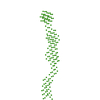
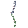
- Structure visualization
| Structure viewer | Molecule: MolmilJmol/JSmol |
|---|
- Downloads & links
-Download
| PDBx/mmCIF format | 5hmo.cif.gz | 111.2 KB | Display | PDBx/mmCIF format |
|---|---|---|---|---|
| PDB format | pdb5hmo.ent.gz | 87.8 KB | Display | PDB format |
| PDBx/mmJSON format | 5hmo.json.gz | Tree view | PDBx/mmJSON format | |
| Others |  Other downloads Other downloads |
-Validation report
| Arichive directory | https://data.pdbj.org/pub/pdb/validation_reports/hm/5hmoftp://data.pdbj.org/pub/pdb/validation_reports/hm/5hmo | HTTPS FTP |
|---|
-Related structure data
| Related structure data | 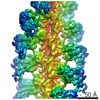 8244C  5hmpC  5i0hC  5i0iC 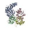 5kg8C  2lw9S S: Starting model for refinement C: citing same article ( |
|---|---|
| Similar structure data |

-Links
 PDBj
PDBj


- Assembly
| Deposited unit | 
| ||||||||
|---|---|---|---|---|---|---|---|---|---|
| 1 |
| ||||||||
| Unit cell |
|
-Components
| #1: Protein | Mass: 16684.678 Da / Num. of mol.: 2 / Fragment: SAH domain and coiled-coil, UNP residues 797-926 Source method: isolated from a genetically manipulated source Source: (gene. exp.)  |
|---|
-Experimental details
-Experiment
| Experiment | Method: X-RAY DIFFRACTION / Number of used crystals: 1 |
|---|
- Sample preparation
| Crystal | Density Matthews: 4.45 Å3/Da / Density % sol: 72.36 % |
|---|---|
| Crystal grow | Temperature: 290 K / Method: vapor diffusion, hanging drop / pH: 7 / Details: PEG 3350, Ammonium citrate, dioxane |
-Data collection
| Diffraction | Mean temperature: 100 K |
|---|---|
| Diffraction source | Source: SYNCHROTRON / Site: SOLEIL / Beamline: PROXIMA 1 / Wavelength: 0.98011 Å |
| Detector | Type: DECTRIS PILATUS 6M / Detector: PIXEL / Date: Jul 6, 2012 |
| Radiation | Protocol: SINGLE WAVELENGTH / Monochromatic (M) / Laue (L): M / Scattering type: x-ray |
| Radiation wavelength | Wavelength: 0.98011 Å / Relative weight: 1 |
| Reflection | Resolution: 3.49→49.37 Å / Num. obs: 7979 / % possible obs: 99.8 % / Redundancy: 13.7 % / Rsym value: 0.1076 / Net I/σ(I): 15.74 |
| Reflection shell | Resolution: 3.49→3.7 Å / Redundancy: 2.08 % / Mean I/σ(I) obs: 2.02 / Rsym value: 1.819 / % possible all: 98.9 |
- Processing
| Software |
| |||||||||||||||||||||||||||||||||||||||||||||||||||||||||||||||||||||||||||
|---|---|---|---|---|---|---|---|---|---|---|---|---|---|---|---|---|---|---|---|---|---|---|---|---|---|---|---|---|---|---|---|---|---|---|---|---|---|---|---|---|---|---|---|---|---|---|---|---|---|---|---|---|---|---|---|---|---|---|---|---|---|---|---|---|---|---|---|---|---|---|---|---|---|---|---|---|
| Refinement | Method to determine structure: MOLECULAR REPLACEMENT Starting model: 2LW9 Resolution: 3.493→45.5 Å / SU ML: 0.55 / Cross valid method: FREE R-VALUE / σ(F): 1.36 / Phase error: 56.04 / Stereochemistry target values: ML
| |||||||||||||||||||||||||||||||||||||||||||||||||||||||||||||||||||||||||||
| Solvent computation | Shrinkage radii: 0.9 Å / VDW probe radii: 1.11 Å / Solvent model: FLAT BULK SOLVENT MODEL | |||||||||||||||||||||||||||||||||||||||||||||||||||||||||||||||||||||||||||
| Displacement parameters | Biso max: 255.39 Å2 / Biso mean: 193.0521 Å2 / Biso min: 123.55 Å2 | |||||||||||||||||||||||||||||||||||||||||||||||||||||||||||||||||||||||||||
| Refinement step | Cycle: final / Resolution: 3.493→45.5 Å
| |||||||||||||||||||||||||||||||||||||||||||||||||||||||||||||||||||||||||||
| Refine LS restraints |
| |||||||||||||||||||||||||||||||||||||||||||||||||||||||||||||||||||||||||||
| LS refinement shell | Refine-ID: X-RAY DIFFRACTION / Total num. of bins used: 3 / % reflection obs: 99 %
| |||||||||||||||||||||||||||||||||||||||||||||||||||||||||||||||||||||||||||
| Refinement TLS params. | Method: refined / Refine-ID: X-RAY DIFFRACTION
| |||||||||||||||||||||||||||||||||||||||||||||||||||||||||||||||||||||||||||
| Refinement TLS group |
|
