 Movie
Movie Controller

[English] 日本語
 Yorodumi
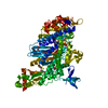
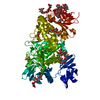
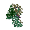
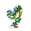
- PDB-4pk4: Myosin VI motor domain in the PPS state - from a Pi release state... -
+ Open data

- Basic information
| Entry | Database: PDB / ID: 4pk4 | ||||||||||||
|---|---|---|---|---|---|---|---|---|---|---|---|---|---|
| Title | Myosin VI motor domain in the PPS state - from a Pi release state crystal, space group P212121 after long soaking with PO4 | ||||||||||||
 Components Components | Unconventional myosin-VI | ||||||||||||
 Keywords Keywords | MOTOR PROTEIN / motor domain / Pre powerstroke state | ||||||||||||
| Function / homology |  Function and homology information Function and homology informationmyosin complex / clathrin-coated vesicle / microvillus / cytoskeletal motor activity / clathrin-coated pit / filopodium / sensory perception of sound / ruffle membrane / endocytosis / actin filament binding ...myosin complex / clathrin-coated vesicle / microvillus / cytoskeletal motor activity / clathrin-coated pit / filopodium / sensory perception of sound / ruffle membrane / endocytosis / actin filament binding / protein transport / calmodulin binding / perinuclear region of cytoplasm / Golgi apparatus / ATP binding / metal ion binding / nucleus / cytosol Similarity search - Function | ||||||||||||
| Biological species |  | ||||||||||||
| Method |  X-RAY DIFFRACTION / SYNCHROTRON / MOLECULAR REPLACEMENT / Resolution: 2.78 Å X-RAY DIFFRACTION / SYNCHROTRON / MOLECULAR REPLACEMENT / Resolution: 2.78 Å | ||||||||||||
 Authors Authors | Isabet, T. / Benisty, H. / Llinas, P. / Sweeney, H.L. / Houdusse, A. | ||||||||||||
| Funding support |  France, France,  United States, 3items United States, 3items
| ||||||||||||
 Citation Citation | Journal: Dev.Cell / Year: 2015 Title: How actin initiates the motor activity of Myosin. Authors: Llinas, P. / Isabet, T. / Song, L. / Ropars, V. / Zong, B. / Benisty, H. / Sirigu, S. / Morris, C. / Kikuti, C. / Safer, D. / Sweeney, H.L. / Houdusse, A. | ||||||||||||
| History |
|
- Structure visualization
| Structure viewer | Molecule: MolmilJmol/JSmol |
|---|
- Downloads & links
-Download
| PDBx/mmCIF format | 4pk4.cif.gz | 324 KB | Display | PDBx/mmCIF format |
|---|---|---|---|---|
| PDB format | pdb4pk4.ent.gz | 262.4 KB | Display | PDB format |
| PDBx/mmJSON format | 4pk4.json.gz | Tree view | PDBx/mmJSON format | |
| Others |  Other downloads Other downloads |
-Validation report
| Arichive directory | https://data.pdbj.org/pub/pdb/validation_reports/pk/4pk4ftp://data.pdbj.org/pub/pdb/validation_reports/pk/4pk4 | HTTPS FTP |
|---|
-Related structure data
| Related structure data |  4pfoC 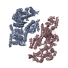 4pfpC 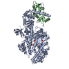 4pjjC 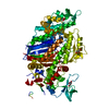 4pjkC 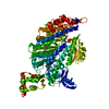 4pjlC 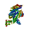 4pjmC  4pjnC 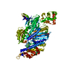 2v26S C: citing same article ( S: Starting model for refinement |
|---|---|
| Similar structure data |


-Links
 PDBj
PDBj


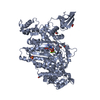
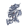
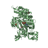
- Assembly
| Deposited unit | 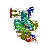
| ||||||||
|---|---|---|---|---|---|---|---|---|---|
| 1 |
| ||||||||
| Unit cell |
|
-Components
| #1: Protein | Mass: 89847.180 Da / Num. of mol.: 1 / Fragment: residues 2-789 Source method: isolated from a genetically manipulated source Source: (gene. exp.)   Spodoptera frugiperda (fall armyworm) / References: UniProt: F1RQI7 Spodoptera frugiperda (fall armyworm) / References: UniProt: F1RQI7 |
|---|---|
| #2: Chemical | ChemComp-ADP /   Mass: 427.201 Da / Num. of mol.: 1 / Source method: obtained synthetically / Formula: C10H15N5O10P2 / Comment: ADP, energy-carrying molecule*YM Mass: 427.201 Da / Num. of mol.: 1 / Source method: obtained synthetically / Formula: C10H15N5O10P2 / Comment: ADP, energy-carrying molecule*YM |
| #3: Chemical | ChemComp-MG /   Mass: 24.305 Da / Num. of mol.: 1 / Source method: obtained synthetically / Formula: Mg Mass: 24.305 Da / Num. of mol.: 1 / Source method: obtained synthetically / Formula: Mg |
| #4: Chemical | ChemComp-PO4 /   Mass: 94.971 Da / Num. of mol.: 1 / Source method: obtained synthetically / Formula: PO4 Mass: 94.971 Da / Num. of mol.: 1 / Source method: obtained synthetically / Formula: PO4 |
| #5: Water | ChemComp-HOH /  Mass: 18.015 Da / Num. of mol.: 15 / Source method: isolated from a natural source / Formula: H2O Mass: 18.015 Da / Num. of mol.: 15 / Source method: isolated from a natural source / Formula: H2O |
-Experimental details
-Experiment
| Experiment | Method: X-RAY DIFFRACTION |
|---|
- Sample preparation
| Crystal | Density Matthews: 2.52 Å3/Da / Density % sol: 51.12 % |
|---|---|
| Crystal grow | Temperature: 277 K / Method: vapor diffusion, hanging drop / pH: 8.5 Details: 6,6% PEG8k, 50mM Tris pH8,5, 1mM TCEP, 3% Glycerol then soaked during 45 minutes with 100mM NaH2PO4 pH7,5 PH range: 8.5 |
-Data collection
| Diffraction | Mean temperature: 100 K |
|---|---|
| Diffraction source | Source: SYNCHROTRON / Site: SOLEIL / Beamline: PROXIMA 1 / Wavelength: 0.9801 Å |
| Detector | Type: ADSC QUANTUM 315r / Detector: CCD / Date: Jul 6, 2011 |
| Radiation | Monochromator: Si III / Protocol: SINGLE WAVELENGTH / Monochromatic (M) / Laue (L): M / Scattering type: x-ray |
| Radiation wavelength | Wavelength: 0.9801 Å / Relative weight: 1 |
| Reflection | Resolution: 2.78→50 Å / Num. obs: 23205 / % possible obs: 99.3 % / Redundancy: 7.23 % / Biso Wilson estimate: 84.54 Å2 / Rmerge(I) obs: 0.127 / Net I/σ(I): 16.53 |
| Reflection shell | Resolution: 2.78→2.85 Å / Redundancy: 4.9 % / Rmerge(I) obs: 1.125 / Mean I/σ(I) obs: 1.35 / % possible all: 98.1 |
- Processing
| Software |
| ||||||||||||||||||||||||||||||||||||||||||||||||||||||||||||||||||||||||||||||||||||||||||||||||||||||||||||||||||
|---|---|---|---|---|---|---|---|---|---|---|---|---|---|---|---|---|---|---|---|---|---|---|---|---|---|---|---|---|---|---|---|---|---|---|---|---|---|---|---|---|---|---|---|---|---|---|---|---|---|---|---|---|---|---|---|---|---|---|---|---|---|---|---|---|---|---|---|---|---|---|---|---|---|---|---|---|---|---|---|---|---|---|---|---|---|---|---|---|---|---|---|---|---|---|---|---|---|---|---|---|---|---|---|---|---|---|---|---|---|---|---|---|---|---|---|
| Refinement | Method to determine structure: MOLECULAR REPLACEMENT Starting model: 2v26 Resolution: 2.78→31 Å / Cor.coef. Fo:Fc: 0.9477 / Cor.coef. Fo:Fc free: 0.8992 / Cross valid method: THROUGHOUT / σ(F): 0 / SU Rfree Blow DPI: 0.361
| ||||||||||||||||||||||||||||||||||||||||||||||||||||||||||||||||||||||||||||||||||||||||||||||||||||||||||||||||||
| Displacement parameters | Biso mean: 104.44 Å2
| ||||||||||||||||||||||||||||||||||||||||||||||||||||||||||||||||||||||||||||||||||||||||||||||||||||||||||||||||||
| Refine analyze | Luzzati coordinate error obs: 0.53 Å | ||||||||||||||||||||||||||||||||||||||||||||||||||||||||||||||||||||||||||||||||||||||||||||||||||||||||||||||||||
| Refinement step | Cycle: 1 / Resolution: 2.78→31 Å
| ||||||||||||||||||||||||||||||||||||||||||||||||||||||||||||||||||||||||||||||||||||||||||||||||||||||||||||||||||
| Refine LS restraints |
| ||||||||||||||||||||||||||||||||||||||||||||||||||||||||||||||||||||||||||||||||||||||||||||||||||||||||||||||||||
| LS refinement shell | Resolution: 2.78→2.9 Å / Total num. of bins used: 12
| ||||||||||||||||||||||||||||||||||||||||||||||||||||||||||||||||||||||||||||||||||||||||||||||||||||||||||||||||||
| Refinement TLS params. | Method: refined / Origin x: 18.9055 Å / Origin y: -21.9872 Å / Origin z: -10.6339 Å
| ||||||||||||||||||||||||||||||||||||||||||||||||||||||||||||||||||||||||||||||||||||||||||||||||||||||||||||||||||
| Refinement TLS group | Selection details: { A|* } |
