- PDB-2v26: Myosin VI (MD) pre-powerstroke state (Mg.ADP.VO4) -
+
Open data
ID or keywords:
Loading...
-
Basic information
Entry
Database: PDB / ID: 2v26
Title
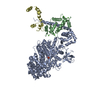
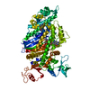
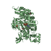
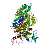
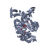
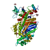
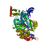
Myosin VI (MD) pre-powerstroke state (Mg.ADP.VO4)
Components
MYOSIN VI
Keywords
STRUCTURAL PROTEIN / CALMODULIN-BINDING / NUCLEOTIDE-BINDING / MYOSIN / MEMBRANE / VANADATE / MYOSIN VI / TRANSPORT / PRE- POWERSTROKE / TRANSITION STATE / PROTEIN TRANSPORT / ACTIN-BINDING / MOTOR PROTEIN / NUCLEAR PROTEIN / ENDOCYTOSIS / ATP-BINDING / COILED COIL / DOMAIN MOTOR / GOLGI APPARATUS / PHOSPHORYLATION / MOLECULAR MOTOR
Function / homology
Function and homology information
regulation of secretion / inner ear auditory receptor cell differentiation / actin filament-based movement / myosin complex / clathrin-coated vesicle / inner ear morphogenesis / microfilament motor activity / filamentous actin / microvillus / cytoskeletal motor activity ...regulation of secretion / inner ear auditory receptor cell differentiation / actin filament-based movement / myosin complex / clathrin-coated vesicle / inner ear morphogenesis / microfilament motor activity / filamentous actin / microvillus / cytoskeletal motor activity / ruffle / actin filament organization / filopodium / actin filament / DNA damage response, signal transduction by p53 class mediator / sensory perception of sound / intracellular protein transport / ADP binding / ruffle membrane / endocytosis / actin filament binding / intracellular protein localization / actin cytoskeleton / cytoplasmic vesicle / nuclear membrane / cell cortex / calmodulin binding / perinuclear region of cytoplasm / Golgi apparatus / nucleoplasm / ATP binding / nucleus / plasma membrane / cytoplasm / cytosol Similarity search - Function
Myosin VI, cargo binding domain / Class VI myosin, motor domain / : / Myosin VI cargo binding domain / Myosin VI, lever arm / Myosin VI head, motor domain, U50 subdomain / Myosin S1 fragment, N-terminal / Methane Monooxygenase Hydroxylase; Chain G, domain 1 - #530 / Kinesin motor domain / Kinesin ...Myosin VI, cargo binding domain / Class VI myosin, motor domain / : / Myosin VI cargo binding domain / Myosin VI, lever arm / Myosin VI head, motor domain, U50 subdomain / Myosin S1 fragment, N-terminal / Methane Monooxygenase Hydroxylase; Chain G, domain 1 - #530 / Kinesin motor domain / Kinesin / Myosin S1 fragment, N-terminal / Myosin, N-terminal, SH3-like / Myosin N-terminal SH3-like domain profile. / Myosin motor domain profile. / Myosin head, motor domain / Myosin head (motor domain) / Myosin. Large ATPases. / Kinesin motor domain superfamily / Methane Monooxygenase Hydroxylase; Chain G, domain 1 / Four Helix Bundle (Hemerythrin (Met), subunit A) / SH3 type barrels. / Roll / Up-down Bundle / P-loop containing nucleoside triphosphate hydrolase / 3-Layer(aba) Sandwich / Mainly Beta / Mainly Alpha / Alpha Beta Similarity search - Domain/homology
Mass: 18.015 Da / Num. of mol.: 536 / Source method: isolated from a natural source / Formula: H2O
-
Details
Sequence details
THE AUTHORS STATE THAT THE ORIGINAL SEQUENCE (UNIPROT Q29122) OF MYOSIN VI FROM PIG WAS MOST LIKELY ...THE AUTHORS STATE THAT THE ORIGINAL SEQUENCE (UNIPROT Q29122) OF MYOSIN VI FROM PIG WAS MOST LIKELY INCORRECT BECAUSE THE CHANGES THAT ARE IN THEIR CLONE (LYS DELETION AND THE 6 MUTATIONS) ARE CONSERVED ACROSS THE MYOSIN VI FAMILY.
-
Experimental details
-
Experiment
Experiment
Method: X-RAY DIFFRACTION
-
Sample preparation
Crystal
Density Matthews: 2.37 Å3/Da / Density % sol: 42.8 % / Description: NONE
Resolution: 1.75→68.68 Å / Cor.coef. Fo:Fc: 0.943 / Cor.coef. Fo:Fc free: 0.93 / SU B: 2.122 / SU ML: 0.071 / Cross valid method: THROUGHOUT / ESU R: 0.128 / ESU R Free: 0.118 / Stereochemistry target values: MAXIMUM LIKELIHOOD / Details: HYDROGENS HAVE BEEN ADDED IN THE RIDING POSITIONS.
Rfactor
Num. reflection
% reflection
Selection details
Rfree
0.227
9071
10.1 %
RANDOM
Rwork
0.202
-
-
-
obs
0.205
81037
99.9 %
-
Solvent computation
Ion probe radii: 0.8 Å / Shrinkage radii: 0.8 Å / VDW probe radii: 1.4 Å / Solvent model: MASK
 Movie
Movie Controller


 Open data
 Basic information
 Components
 Keywords
 Function and homology information
 X-RAY DIFFRACTION /
  Authors
 Citation
 Structure visualization
 Downloads & links
 Other downloads
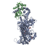


 PDBj
PDBj


 Assembly
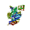

 SPODOPTERA FRUGIPERDA (fall armyworm) / References: UniProt: Q29122


 Mass: 427.201 Da / Num. of mol.: 1 / Source method: obtained synthetically / Formula: C10H15N5O10P2 / Comment: ADP, energy-carrying molecule*YM
 Mass: 114.939 Da / Num. of mol.: 1 / Source method: obtained synthetically / Formula: VO4
 Mass: 62.068 Da / Num. of mol.: 7 / Source method: obtained synthetically / Formula: C2H6O2
 Mass: 96.063 Da / Num. of mol.: 2 / Source method: obtained synthetically / Formula: SO4
 Mass: 24.305 Da / Num. of mol.: 1 / Source method: obtained synthetically / Formula: Mg
 Sample preparation
 / Beamline: ID23-1 / Wavelength: 1.008
  Processing