 Movie
Movie Controller


+ Open data

- Basic information
| Entry | Database: PDB / ID: 2bkh | ||||||
|---|---|---|---|---|---|---|---|
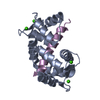
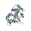
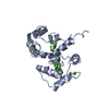
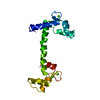
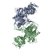
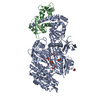
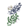
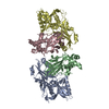
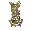
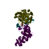
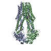
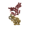
| Title | Myosin VI nucleotide-free (MDInsert2) crystal structure | ||||||
 Components Components |
| ||||||
 Keywords Keywords | MOTOR PROTEIN/METAL-BINDING PROTEIN / COMPLEX (MOTOR PROTEIN-CALMODULIN) / MYOSIN VI / REVERSE MYOSIN / CALMODULIN / NON-CONVENTIONAL MYOSIN / NUCLEOTIDE-FREE CONFORMATION / MUSCLE PROTEIN / MOTOR PROTEIN-METAL-BINDING PROTEIN complex | ||||||
| Function / homology |  Function and homology information Function and homology informationnegative regulation of phospholipase C-activating phototransduction signaling pathway / myosin VI complex / myosin VI head/neck binding / myosin VII complex / photoreceptor cell axon guidance / negative regulation of opsin-mediated signaling pathway / rhabdomere development / rhabdomere / myosin V complex / regulation of secretion ...negative regulation of phospholipase C-activating phototransduction signaling pathway / myosin VI complex / myosin VI head/neck binding / myosin VII complex / photoreceptor cell axon guidance / negative regulation of opsin-mediated signaling pathway / rhabdomere development / rhabdomere / myosin V complex / regulation of secretion / detection of chemical stimulus involved in sensory perception of smell / kinetochore organization / autophagic cell death / G protein-coupled opsin signaling pathway / inner ear auditory receptor cell differentiation / actin filament-based movement / myosin V binding / channel regulator activity / myosin complex / clathrin-coated vesicle / inner ear morphogenesis / muscle cell cellular homeostasis / mitotic spindle pole / microfilament motor activity / myosin heavy chain binding / centriole replication / filamentous actin / microvillus / cytoskeletal motor activity / cellular response to ethanol / enzyme regulator activity / ruffle / actin filament organization / filopodium / actin filament / DNA damage response, signal transduction by p53 class mediator / centriole / sensory perception of sound / ADP binding / intracellular protein transport / microtubule cytoskeleton organization / spindle / ruffle membrane / endocytosis / actin filament binding / sensory perception of smell / mitotic spindle / intracellular protein localization / actin cytoskeleton / cytoplasmic vesicle / midbody / cell cortex / nuclear membrane / calmodulin binding / calcium ion binding / centrosome / perinuclear region of cytoplasm / Golgi apparatus / nucleoplasm / ATP binding / nucleus / plasma membrane / cytoplasm / cytosol Similarity search - Function | ||||||
| Biological species |   | ||||||
| Method |  X-RAY DIFFRACTION / SYNCHROTRON / MOLECULAR REPLACEMENT / Resolution: 2.4 Å X-RAY DIFFRACTION / SYNCHROTRON / MOLECULAR REPLACEMENT / Resolution: 2.4 Å | ||||||
 Authors Authors | Menetrey, J. / Bahloul, A. / Yengo, C. / Wells, A. / Morris, C. / Sweeney, H.L. / Houdusse, A. | ||||||
 Citation Citation | Journal: Nature / Year: 2005 Title: The Structure of the Myosin Vi Motor Reveals the Mechanism of Directionality Reversal Authors: Menetrey, J. / Bahloul, A. / Wells, A. / Yengo, C. / Morris, C. / Sweeney, H.L. / Houdusse, A. | ||||||
| History |
|
- Structure visualization
| Structure viewer | Molecule: MolmilJmol/JSmol |
|---|
- Downloads & links
-Download
| PDBx/mmCIF format | 2bkh.cif.gz | 204.8 KB | Display | PDBx/mmCIF format |
|---|---|---|---|---|
| PDB format | pdb2bkh.ent.gz | 159.9 KB | Display | PDB format |
| PDBx/mmJSON format | 2bkh.json.gz | Tree view | PDBx/mmJSON format | |
| Others |  Other downloads Other downloads |
-Validation report
| Arichive directory | https://data.pdbj.org/pub/pdb/validation_reports/bk/2bkhftp://data.pdbj.org/pub/pdb/validation_reports/bk/2bkh | HTTPS FTP |
|---|
-Related structure data
| Related structure data | 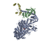 2bkiC 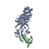 1oe9S C: citing same article ( S: Starting model for refinement |
|---|---|
| Similar structure data |


-Links
 PDBj
PDBj


- Assembly
| Deposited unit | 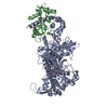
| ||||||||
|---|---|---|---|---|---|---|---|---|---|
| 1 |
| ||||||||
| Unit cell |
|
-Components
| #1: Protein | Mass: 93033.078 Da / Num. of mol.: 1 / Fragment: MOTOR DOMAIN-INSERT2, RESIDUES 2-816 Source method: isolated from a genetically manipulated source Source: (gene. exp.)  SPODOPTERA FRUGIPERDA (fall armyworm) / References: UniProt: Q29122 SPODOPTERA FRUGIPERDA (fall armyworm) / References: UniProt: Q29122 | ||||||
|---|---|---|---|---|---|---|---|
| #2: Protein | Mass: 16825.520 Da / Num. of mol.: 1 Source method: isolated from a genetically manipulated source Source: (gene. exp.) SPODOPTERA FRUGIPERDA (fall armyworm) / References: UniProt: P62152 | ||||||
| #3: Chemical | ChemComp-GOL /   Mass: 92.094 Da / Num. of mol.: 6 / Source method: obtained synthetically / Formula: C3H8O3 Mass: 92.094 Da / Num. of mol.: 6 / Source method: obtained synthetically / Formula: C3H8O3#4: Chemical | ChemComp-CA /   Mass: 40.078 Da / Num. of mol.: 4 / Source method: obtained synthetically / Formula: Ca Mass: 40.078 Da / Num. of mol.: 4 / Source method: obtained synthetically / Formula: Ca#5: Water | ChemComp-HOH / |  Mass: 18.015 Da / Num. of mol.: 285 / Source method: isolated from a natural source / Formula: H2O Mass: 18.015 Da / Num. of mol.: 285 / Source method: isolated from a natural source / Formula: H2OSequence details | THE AUTHORS STATE THAT THE ORIGINAL SEQUENCE (UNIPROT Q29122) OF MYOSIN VI FROM PIG WAS MOST LIKELY ...THE AUTHORS STATE THAT THE ORIGINAL SEQUENCE (UNIPROT Q29122) OF MYOSIN VI FROM PIG WAS MOST LIKELY INCORRECT BECAUSE THE CHANGES THAT ARE IN THEIR CLONE (LYS DELETION AND THE 6 MUTATIONS) ARE CONSERVED ACROSS THE MYOSIN VI FAMILY. | |
-Experimental details
-Experiment
| Experiment | Method: X-RAY DIFFRACTION |
|---|
- Sample preparation
| Crystal | Density Matthews: 2.99 Å3/Da / Density % sol: 58.5 % |
|---|---|
| Crystal grow | Details: 8-10% PEG 8000, 50MM MES PH 6.7, 150MM NH4.SO4, 3% ISO-PROPANOL, 3% TERT-BUTANOL |
-Data collection
| Diffraction | Mean temperature: 100 K |
|---|---|
| Diffraction source | Source: SYNCHROTRON / Site: ESRF  / Beamline: ID14-1 / Wavelength: 0.934 / Beamline: ID14-1 / Wavelength: 0.934 |
| Detector | Type: ADSC CCD / Detector: CCD |
| Radiation | Protocol: SINGLE WAVELENGTH / Monochromatic (M) / Laue (L): M / Scattering type: x-ray |
| Radiation wavelength | Wavelength: 0.934 Å / Relative weight: 1 |
| Reflection | Resolution: 2.4→40 Å / Num. obs: 49721 / % possible obs: 99.8 % / Observed criterion σ(I): 2 / Redundancy: 4 % / Rmerge(I) obs: 0.09 / Net I/σ(I): 14.4 |
| Reflection shell | Resolution: 2.4→2.49 Å / Rmerge(I) obs: 0.42 / Mean I/σ(I) obs: 3.2 / % possible all: 99.9 |
- Processing
| Software |
| ||||||||||||||||||||||||||||||||||||||||||||||||||||||||||||||||||||||||||||||||||||||||||||||||||||||||||||||||||||||||||||||||||||||||||||||||||||||||||||||||||||||||||||||||||||||
|---|---|---|---|---|---|---|---|---|---|---|---|---|---|---|---|---|---|---|---|---|---|---|---|---|---|---|---|---|---|---|---|---|---|---|---|---|---|---|---|---|---|---|---|---|---|---|---|---|---|---|---|---|---|---|---|---|---|---|---|---|---|---|---|---|---|---|---|---|---|---|---|---|---|---|---|---|---|---|---|---|---|---|---|---|---|---|---|---|---|---|---|---|---|---|---|---|---|---|---|---|---|---|---|---|---|---|---|---|---|---|---|---|---|---|---|---|---|---|---|---|---|---|---|---|---|---|---|---|---|---|---|---|---|---|---|---|---|---|---|---|---|---|---|---|---|---|---|---|---|---|---|---|---|---|---|---|---|---|---|---|---|---|---|---|---|---|---|---|---|---|---|---|---|---|---|---|---|---|---|---|---|---|---|
| Refinement | Method to determine structure: MOLECULAR REPLACEMENT Starting model: PDB ENTRY 1OE9 Resolution: 2.4→40 Å / Cor.coef. Fo:Fc: 0.942 / Cor.coef. Fo:Fc free: 0.906 / SU B: 7.454 / SU ML: 0.174 / Cross valid method: THROUGHOUT / ESU R: 0.347 / ESU R Free: 0.251 / Stereochemistry target values: MAXIMUM LIKELIHOOD Details: HYDROGENS HAVE BEEN ADDED IN THE RIDING POSITIONS. DISORDERED REGIONS 356-360 AND 623-639 IN CHAIN A AND 73-80 IN CHAIN B WERE NOT MODELED.
| ||||||||||||||||||||||||||||||||||||||||||||||||||||||||||||||||||||||||||||||||||||||||||||||||||||||||||||||||||||||||||||||||||||||||||||||||||||||||||||||||||||||||||||||||||||||
| Solvent computation | Ion probe radii: 0.8 Å / Shrinkage radii: 0.8 Å / VDW probe radii: 1.4 Å / Solvent model: BABINET MODEL WITH MASK | ||||||||||||||||||||||||||||||||||||||||||||||||||||||||||||||||||||||||||||||||||||||||||||||||||||||||||||||||||||||||||||||||||||||||||||||||||||||||||||||||||||||||||||||||||||||
| Displacement parameters | Biso mean: 45.1 Å2
| ||||||||||||||||||||||||||||||||||||||||||||||||||||||||||||||||||||||||||||||||||||||||||||||||||||||||||||||||||||||||||||||||||||||||||||||||||||||||||||||||||||||||||||||||||||||
| Refinement step | Cycle: LAST / Resolution: 2.4→40 Å
| ||||||||||||||||||||||||||||||||||||||||||||||||||||||||||||||||||||||||||||||||||||||||||||||||||||||||||||||||||||||||||||||||||||||||||||||||||||||||||||||||||||||||||||||||||||||
| Refine LS restraints |
|