 Movie
Movie Controller

[English] 日本語
 Yorodumi
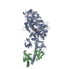
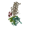
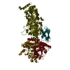
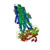
- PDB-1oe9: Crystal structure of Myosin V motor with essential light chain-nu... -
+ Open data

- Basic information
| Entry | Database: PDB / ID: 1oe9 | ||||||
|---|---|---|---|---|---|---|---|
| Title | Crystal structure of Myosin V motor with essential light chain-nucleotide-free | ||||||
 Components Components |
| ||||||
 Keywords Keywords | ATPASE/MYOSIN / ATPASE-MYOSIN COMPLEX / UNCONVENTIONAL MYOSIN / MYOSIN V / CHICKEN / MOLECULAR MOTOR / ATPASE / ELC / IQ MOTIF / MUSCLE PROTEIN / ATP-BINDING | ||||||
| Function / homology |  Function and homology information Function and homology informationminus-end directed microfilament motor activity / insulin-responsive compartment / unconventional myosin complex / muscle myosin complex / muscle filament sliding / myosin complex / myosin II complex / structural constituent of muscle / microfilament motor activity / filamentous actin ...minus-end directed microfilament motor activity / insulin-responsive compartment / unconventional myosin complex / muscle myosin complex / muscle filament sliding / myosin complex / myosin II complex / structural constituent of muscle / microfilament motor activity / filamentous actin / cytoskeletal motor activity / Smooth Muscle Contraction / skeletal muscle tissue development / vesicle-mediated transport / muscle contraction / actin filament organization / protein localization to plasma membrane / cellular response to insulin stimulus / endocytosis / actin filament binding / actin cytoskeleton / calmodulin binding / Golgi membrane / calcium ion binding / ATP hydrolysis activity / extracellular exosome / ATP binding / membrane / cytoplasm / cytosol Similarity search - Function | ||||||
| Biological species |   HOMO SAPIENS (human) HOMO SAPIENS (human) | ||||||
| Method |  X-RAY DIFFRACTION / SYNCHROTRON / MOLECULAR REPLACEMENT / Resolution: 2.05 Å X-RAY DIFFRACTION / SYNCHROTRON / MOLECULAR REPLACEMENT / Resolution: 2.05 Å | ||||||
 Authors Authors | Coureux, P.-D. / Wells, A.L. / Menetrey, J. / Yengo, C.M. / Morris, C.A. / Sweeney, H.L. / Houdusse, A. | ||||||
 Citation Citation | Journal: Nature / Year: 2003 Title: A Structural State of the Myosin V Motor without Bound Nucleotide Authors: Coureux, P.-D. / Wells, A.L. / Menetrey, J. / Yengo, C.M. / Morris, C.A. / Sweeney, H.L. / Houdusse, A. | ||||||
| History |
|
- Structure visualization
| Structure viewer | Molecule: MolmilJmol/JSmol |
|---|
- Downloads & links
-Download
| PDBx/mmCIF format | 1oe9.cif.gz | 192.6 KB | Display | PDBx/mmCIF format |
|---|---|---|---|---|
| PDB format | pdb1oe9.ent.gz | 150.6 KB | Display | PDB format |
| PDBx/mmJSON format | 1oe9.json.gz | Tree view | PDBx/mmJSON format | |
| Others |  Other downloads Other downloads |
-Validation report
| Arichive directory | https://data.pdbj.org/pub/pdb/validation_reports/oe/1oe9ftp://data.pdbj.org/pub/pdb/validation_reports/oe/1oe9 | HTTPS FTP |
|---|
-Related structure data
| Related structure data | 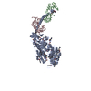 2mysS S: Starting model for refinement |
|---|---|
| Similar structure data |


-Links
 PDBj
PDBj


- Assembly
| Deposited unit | 
| ||||||||
|---|---|---|---|---|---|---|---|---|---|
| 1 |
| ||||||||
| Unit cell |
|
-Components
| #1: Protein | Mass: 91607.172 Da / Num. of mol.: 1 / Fragment: MOTOR DOMAIN, RESIDUES 1-792 Source method: isolated from a genetically manipulated source Source: (gene. exp.)   SPODOPTERA FRUGIPERDA (fall armyworm) / References: UniProt: Q02440 SPODOPTERA FRUGIPERDA (fall armyworm) / References: UniProt: Q02440 |
|---|---|
| #2: Protein | Mass: 17090.277 Da / Num. of mol.: 1 / Fragment: RESIDUES 59-208 Source method: isolated from a genetically manipulated source Source: (gene. exp.) HOMO SAPIENS (human) / Cell line (production host): SF9 / Production host: SPODOPTERA FRUGIPERDA (fall armyworm) / References: UniProt: P14649 |
| #3: Chemical | ChemComp-SO4 /   Mass: 96.063 Da / Num. of mol.: 1 / Source method: obtained synthetically / Formula: SO4 Mass: 96.063 Da / Num. of mol.: 1 / Source method: obtained synthetically / Formula: SO4 |
| #4: Water | ChemComp-HOH /  Mass: 18.015 Da / Num. of mol.: 333 / Source method: isolated from a natural source / Formula: H2O Mass: 18.015 Da / Num. of mol.: 333 / Source method: isolated from a natural source / Formula: H2O |
| Compound details | MYOSIN VA: PROCESSIVE ACTIN-BASED MOTOR THAT CAN MOVE IN LARGE STEPS. POSSIBLY INVOLVED IN ...MYOSIN VA: PROCESSIVE |
| Sequence details | THE LAST THREE RESIDUES AT THE C-TERMINUS FOR CHAIN A DERIVE FROM THE EXPRESSION VECTOR USED IN THE ...THE LAST THREE RESIDUES AT THE C-TERMINUS FOR CHAIN A DERIVE FROM THE EXPRESSION |
-Experimental details
-Experiment
| Experiment | Method: X-RAY DIFFRACTION / Number of used crystals: 1 |
|---|
- Sample preparation
| Crystal | Density Matthews: 2.8 Å3/Da / Density % sol: 47.8 % | ||||||||||||||||||||||||||||||||||||||||||
|---|---|---|---|---|---|---|---|---|---|---|---|---|---|---|---|---|---|---|---|---|---|---|---|---|---|---|---|---|---|---|---|---|---|---|---|---|---|---|---|---|---|---|---|
| Crystal grow | pH: 6.5 Details: 6% PEG8000 (W/V), 50MM MOPS PH 6.5, 2MM DTT, 2MM NAN3 | ||||||||||||||||||||||||||||||||||||||||||
| Crystal grow | *PLUS pH: 6.5 / Method: vapor diffusion, hanging drop | ||||||||||||||||||||||||||||||||||||||||||
| Components of the solutions | *PLUS
|
-Data collection
| Diffraction | Mean temperature: 100 K |
|---|---|
| Diffraction source | Source: SYNCHROTRON / Site: ESRF  / Beamline: ID29 / Wavelength: 0.9756 / Beamline: ID29 / Wavelength: 0.9756 |
| Detector | Date: Nov 15, 2002 |
| Radiation | Protocol: SINGLE WAVELENGTH / Monochromatic (M) / Laue (L): M / Scattering type: x-ray |
| Radiation wavelength | Wavelength: 0.9756 Å / Relative weight: 1 |
| Reflection | Resolution: 2.05→40 Å / Num. obs: 69115 / % possible obs: 96.8 % / Observed criterion σ(I): 3 / Redundancy: 2.9 % / Rmerge(I) obs: 0.06 / Net I/σ(I): 14 |
| Reflection shell | Resolution: 2.05→2.12 Å / Redundancy: 2 % / Rmerge(I) obs: 0.335 / Mean I/σ(I) obs: 2.34 / % possible all: 88.4 |
| Reflection | *PLUS Highest resolution: 2 Å / Rmerge(I) obs: 0.06 |
| Reflection shell | *PLUS % possible obs: 88.4 % / Rmerge(I) obs: 0.335 |
- Processing
| Software |
| ||||||||||||||||||||||||||||||||||||||||||||||||||||||||||||||||||||||||||||||||||||||||||||||||||||||||||||||||||||||||||||||||||||||||||||||||||||||||||||||||||||||||||||||||||||||
|---|---|---|---|---|---|---|---|---|---|---|---|---|---|---|---|---|---|---|---|---|---|---|---|---|---|---|---|---|---|---|---|---|---|---|---|---|---|---|---|---|---|---|---|---|---|---|---|---|---|---|---|---|---|---|---|---|---|---|---|---|---|---|---|---|---|---|---|---|---|---|---|---|---|---|---|---|---|---|---|---|---|---|---|---|---|---|---|---|---|---|---|---|---|---|---|---|---|---|---|---|---|---|---|---|---|---|---|---|---|---|---|---|---|---|---|---|---|---|---|---|---|---|---|---|---|---|---|---|---|---|---|---|---|---|---|---|---|---|---|---|---|---|---|---|---|---|---|---|---|---|---|---|---|---|---|---|---|---|---|---|---|---|---|---|---|---|---|---|---|---|---|---|---|---|---|---|---|---|---|---|---|---|---|
| Refinement | Method to determine structure: MOLECULAR REPLACEMENT Starting model: PDB ENTRY 2MYS Resolution: 2.05→40 Å / Cor.coef. Fo:Fc: 0.94 / Cor.coef. Fo:Fc free: 0.912 / SU B: 4.969 / SU ML: 0.135 / Cross valid method: THROUGHOUT / ESU R: 0.206 / ESU R Free: 0.185 / Stereochemistry target values: MAXIMUM LIKELIHOOD Details: HYDROGENS HAVE BEEN ADDED IN THE RIDING POSITIONS.DISORDERED REGIONS WERE NOT MODELED.DISORDERED SIDE-CHAINS WERE NOT MODELED.
| ||||||||||||||||||||||||||||||||||||||||||||||||||||||||||||||||||||||||||||||||||||||||||||||||||||||||||||||||||||||||||||||||||||||||||||||||||||||||||||||||||||||||||||||||||||||
| Solvent computation | Ion probe radii: 0.8 Å / Shrinkage radii: 0.8 Å / VDW probe radii: 1.4 Å / Solvent model: BABINET MODEL WITH MASK | ||||||||||||||||||||||||||||||||||||||||||||||||||||||||||||||||||||||||||||||||||||||||||||||||||||||||||||||||||||||||||||||||||||||||||||||||||||||||||||||||||||||||||||||||||||||
| Displacement parameters | Biso mean: 39.16 Å2
| ||||||||||||||||||||||||||||||||||||||||||||||||||||||||||||||||||||||||||||||||||||||||||||||||||||||||||||||||||||||||||||||||||||||||||||||||||||||||||||||||||||||||||||||||||||||
| Refinement step | Cycle: LAST / Resolution: 2.05→40 Å
| ||||||||||||||||||||||||||||||||||||||||||||||||||||||||||||||||||||||||||||||||||||||||||||||||||||||||||||||||||||||||||||||||||||||||||||||||||||||||||||||||||||||||||||||||||||||
| Refine LS restraints |
|