 Movie
Movie Controller

[English] 日本語
 Yorodumi
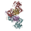
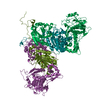
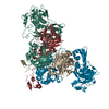
- PDB-7by6: Plasmodium vivax cytoplasmic Phenylalanyl-tRNA synthetase in comp... -
+ Open data

- Basic information
| Entry | Database: PDB / ID: 7by6 | ||||||
|---|---|---|---|---|---|---|---|
| Title | Plasmodium vivax cytoplasmic Phenylalanyl-tRNA synthetase in complex with BRD1389 | ||||||
 Components Components | (Phenylalanyl-tRNA synthetase beta chain, putative) x 2 | ||||||
 Keywords Keywords | LIGASE / AMINOACYLATION / AMINOACYL-TRNA SYNTHETASE / TRNA-BINDING / ATP-BINDING / AUXILIARY POCKET / HETEROTETRAMERIC | ||||||
| Function / homology |  Function and homology information Function and homology informationphenylalanine-tRNA ligase complex / phenylalanine-tRNA ligase / phenylalanyl-tRNA aminoacylation / phenylalanine-tRNA ligase activity / protein heterotetramerization / tRNA binding / magnesium ion binding / RNA binding / ATP binding / cytoplasm / cytosol Similarity search - Function | ||||||
| Biological species |  | ||||||
| Method |  X-RAY DIFFRACTION / SYNCHROTRON / MOLECULAR REPLACEMENT / molecular replacement / Resolution: 2.997 Å X-RAY DIFFRACTION / SYNCHROTRON / MOLECULAR REPLACEMENT / molecular replacement / Resolution: 2.997 Å | ||||||
 Authors Authors | Malhotra, N. / Manmohan, S. / Harlos, K. / Melillo, B. / Schreiber, S.L. / Manickam, Y. / Sharma, S. | ||||||
 Citation Citation | Journal: Nat Commun / Year: 2021 Title: Structural basis of malaria parasite phenylalanine tRNA-synthetase inhibition by bicyclic azetidines. Authors: Sharma, M. / Malhotra, N. / Yogavel, M. / Harlos, K. / Melillo, B. / Comer, E. / Gonse, A. / Parvez, S. / Mitasev, B. / Fang, F.G. / Schreiber, S.L. / Sharma, A. | ||||||
| History |
|
- Structure visualization
| Structure viewer | Molecule: MolmilJmol/JSmol |
|---|
- Downloads & links
-Download
| PDBx/mmCIF format | 7by6.cif.gz | 369.5 KB | Display | PDBx/mmCIF format |
|---|---|---|---|---|
| PDB format | pdb7by6.ent.gz | 297.9 KB | Display | PDB format |
| PDBx/mmJSON format | 7by6.json.gz | Tree view | PDBx/mmJSON format | |
| Others |  Other downloads Other downloads |
-Validation report
| Arichive directory | https://data.pdbj.org/pub/pdb/validation_reports/by/7by6ftp://data.pdbj.org/pub/pdb/validation_reports/by/7by6 | HTTPS FTP |
|---|
-Related structure data
| Related structure data | 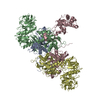 3l4gS S: Starting model for refinement |
|---|---|
| Similar structure data |


-Links
 PDBj
PDBj

- Assembly
| Deposited unit | 
| ||||||||
|---|---|---|---|---|---|---|---|---|---|
| 1 | 
| ||||||||
| Unit cell |
|
-Components
| #1: Protein | Mass: 35323.441 Da / Num. of mol.: 1 Source method: isolated from a genetically manipulated source Source: (gene. exp.) Gene: PVX_081300 / Plasmid: pETM11 / Production host:  |
|---|---|
| #2: Protein | Mass: 71532.250 Da / Num. of mol.: 1 Source method: isolated from a genetically manipulated source Source: (gene. exp.) Gene: PVX_090880 / Plasmid: pETM41 / Production host: |
| #3: Chemical | ChemComp-FB9 / (  Mass: 567.675 Da / Num. of mol.: 1 / Source method: obtained synthetically / Formula: C34H37N3O5 / Feature type: SUBJECT OF INVESTIGATION Mass: 567.675 Da / Num. of mol.: 1 / Source method: obtained synthetically / Formula: C34H37N3O5 / Feature type: SUBJECT OF INVESTIGATION |
| #4: Chemical | ChemComp-MG /   Mass: 24.305 Da / Num. of mol.: 1 / Source method: obtained synthetically / Formula: Mg / Feature type: SUBJECT OF INVESTIGATION Mass: 24.305 Da / Num. of mol.: 1 / Source method: obtained synthetically / Formula: Mg / Feature type: SUBJECT OF INVESTIGATION |
| Has ligand of interest | Y |
-Experimental details
-Experiment
| Experiment | Method: X-RAY DIFFRACTION / Number of used crystals: 1 |
|---|
- Sample preparation
| Crystal | Density Matthews: 2.88 Å3/Da / Density % sol: 57.27 % |
|---|---|
| Crystal grow | Temperature: 293 K / Method: vapor diffusion, hanging drop / pH: 6.5 Details: 0.1 M sodium cacodylate, 3% w/v poly-gama-glutamic acid (Na+ form, low molecular weight), 3% w/v PEG20000, 0.1 M ammonium sulphate, 0.3 M sodium formate |
-Data collection
| Diffraction | Mean temperature: 100 K / Serial crystal experiment: N |
|---|---|
| Diffraction source | Source: SYNCHROTRON / Site: Diamond  / Beamline: I02 / Wavelength: 0.9688 Å / Beamline: I02 / Wavelength: 0.9688 Å |
| Detector | Type: DECTRIS PILATUS3 S 6M / Detector: PIXEL / Date: Apr 12, 2019 |
| Radiation | Protocol: SINGLE WAVELENGTH / Monochromatic (M) / Laue (L): M / Scattering type: x-ray |
| Radiation wavelength | Wavelength: 0.9688 Å / Relative weight: 1 |
| Reflection | Resolution: 2.997→136.5 Å / Num. obs: 25453 / % possible obs: 99.6 % / Redundancy: 6.3 % / CC1/2: 0.921 / Rrim(I) all: 0.145 / Net I/σ(I): 8.5 |
| Reflection shell | Resolution: 3→3.05 Å / Redundancy: 6.3 % / Mean I/σ(I) obs: 1.6 / Num. unique obs: 1186 / CC1/2: 0.741 / Rrim(I) all: 1.288 / % possible all: 94.6 |
-Phasing
| Phasing | Method: molecular replacement |
|---|
- Processing
| Software |
| ||||||||||||||||||||||||||||||||||||||||||||||||||||||||||||
|---|---|---|---|---|---|---|---|---|---|---|---|---|---|---|---|---|---|---|---|---|---|---|---|---|---|---|---|---|---|---|---|---|---|---|---|---|---|---|---|---|---|---|---|---|---|---|---|---|---|---|---|---|---|---|---|---|---|---|---|---|---|
| Refinement | Method to determine structure: MOLECULAR REPLACEMENT Starting model: 3L4G Resolution: 2.997→121.7824 Å / SU ML: 0.44 / Cross valid method: THROUGHOUT / σ(F): 1.33 / Phase error: 32.67 / Stereochemistry target values: ML
| ||||||||||||||||||||||||||||||||||||||||||||||||||||||||||||
| Solvent computation | Shrinkage radii: 0.9 Å / VDW probe radii: 1.11 Å / Solvent model: FLAT BULK SOLVENT MODEL | ||||||||||||||||||||||||||||||||||||||||||||||||||||||||||||
| Displacement parameters | Biso max: 209.44 Å2 / Biso mean: 91.2782 Å2 / Biso min: 47.14 Å2 | ||||||||||||||||||||||||||||||||||||||||||||||||||||||||||||
| Refinement step | Cycle: final / Resolution: 2.997→121.7824 Å
| ||||||||||||||||||||||||||||||||||||||||||||||||||||||||||||
| LS refinement shell | Refine-ID: X-RAY DIFFRACTION / Rfactor Rfree error: 0
| ||||||||||||||||||||||||||||||||||||||||||||||||||||||||||||
| Refinement TLS params. | Method: refined / Origin x: 21.8053 Å / Origin y: 3.5471 Å / Origin z: 25.3067 Å
| ||||||||||||||||||||||||||||||||||||||||||||||||||||||||||||
| Refinement TLS group |
|
