 Movie
Movie Controller

[English] 日本語
 Yorodumi
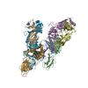
- PDB-3l4g: Crystal structure of Homo Sapiens cytoplasmic Phenylalanyl-tRNA s... -
+ Open data

- Basic information
| Entry | Database: PDB / ID: 3l4g | ||||||
|---|---|---|---|---|---|---|---|
| Title | Crystal structure of Homo Sapiens cytoplasmic Phenylalanyl-tRNA synthetase | ||||||
 Components Components |
| ||||||
 Keywords Keywords | LIGASE / aminoacylation / tRNA-binding / DNA-binding domain / four-helix bundle / Acetylation / Aminoacyl-tRNA synthetase / ATP-binding / Cytoplasm / Nucleotide-binding / Phosphoprotein / Polymorphism / Protein biosynthesis | ||||||
| Function / homology |  Function and homology information Function and homology informationphenylalanine-tRNA ligase complex / phenylalanine-tRNA ligase / phenylalanyl-tRNA aminoacylation / phenylalanine-tRNA ligase activity / Cytosolic tRNA aminoacylation / protein heterotetramerization / tRNA binding / translation / magnesium ion binding / RNA binding ...phenylalanine-tRNA ligase complex / phenylalanine-tRNA ligase / phenylalanyl-tRNA aminoacylation / phenylalanine-tRNA ligase activity / Cytosolic tRNA aminoacylation / protein heterotetramerization / tRNA binding / translation / magnesium ion binding / RNA binding / nucleoplasm / ATP binding / membrane / cytosol / cytoplasm Similarity search - Function | ||||||
| Biological species |  Homo sapiens (human) Homo sapiens (human) | ||||||
| Method |  X-RAY DIFFRACTION / SYNCHROTRON / MOLECULAR REPLACEMENT / Resolution: 3.3 Å X-RAY DIFFRACTION / SYNCHROTRON / MOLECULAR REPLACEMENT / Resolution: 3.3 Å | ||||||
 Authors Authors | Finarov, I. / Moor, N. / Kessler, N. / Klipcan, L. / Safro, M.G. | ||||||
 Citation Citation | Journal: Structure / Year: 2010 Title: Structure of human cytosolic phenylalanyl-tRNA synthetase: evidence for kingdom-specific design of the active sites and tRNA binding patterns. Authors: Finarov, I. / Moor, N. / Kessler, N. / Klipcan, L. / Safro, M.G. #1: Journal: Acta Crystallogr.,Sect.F / Year: 2009Title: Crystallization and X-ray analysis of human cytoplasmic phenylalanyl-tRNA synthetase Authors: Finarov, I. / Moor, N. / Kessler, N. / Safro, M.G. | ||||||
| History |
|
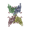
- Structure visualization
| Structure viewer | Molecule: MolmilJmol/JSmol |
|---|
- Downloads & links
-Download
| PDBx/mmCIF format | 3l4g.cif.gz | 1.4 MB | Display | PDBx/mmCIF format |
|---|---|---|---|---|
| PDB format | pdb3l4g.ent.gz | 1.2 MB | Display | PDB format |
| PDBx/mmJSON format | 3l4g.json.gz | Tree view | PDBx/mmJSON format | |
| Others |  Other downloads Other downloads |
-Validation report
| Arichive directory | https://data.pdbj.org/pub/pdb/validation_reports/l4/3l4gftp://data.pdbj.org/pub/pdb/validation_reports/l4/3l4g | HTTPS FTP |
|---|
-Related structure data

-Links
 PDBj
PDBj


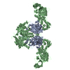
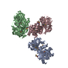
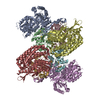
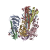
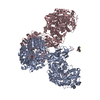
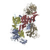
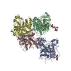
- Assembly
| Deposited unit | 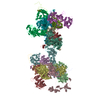
| ||||||||
|---|---|---|---|---|---|---|---|---|---|
| 1 | 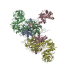
| ||||||||
| 2 | 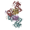
| ||||||||
| 3 | 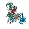
| ||||||||
| 4 | 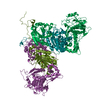
| ||||||||
| Unit cell |
|
-Components
| #1: Protein | Mass: 57644.527 Da / Num. of mol.: 8 Source method: isolated from a genetically manipulated source Source: (gene. exp.) Homo sapiens (human) / Gene: FARSA, FARS, FARSL, FARSLA / Plasmid: Pet21 / Production host:  #2: Protein | Mass: 66214.484 Da / Num. of mol.: 8 Source method: isolated from a genetically manipulated source Source: (gene. exp.) Homo sapiens (human) / Gene: FARSB, FARSLB, FRSB, HSPC173 / Plasmid: PET28 / Production host: #3: Chemical | ChemComp-PHE /   Type: L-peptide linking / Mass: 165.189 Da / Num. of mol.: 8 / Source method: obtained synthetically / Formula: C9H11NO2 Type: L-peptide linking / Mass: 165.189 Da / Num. of mol.: 8 / Source method: obtained synthetically / Formula: C9H11NO2 |
|---|
-Experimental details
-Experiment
| Experiment | Method: X-RAY DIFFRACTION / Number of used crystals: 1 |
|---|
- Sample preparation
| Crystal | Density Matthews: 3.41 Å3/Da / Density % sol: 63.95 % |
|---|---|
| Crystal grow | Temperature: 298 K / Method: vapor diffusion, hanging drop / pH: 6.4 Details: 1.04M NaCitrate, MES pH 6.4, 5mM 2-mercaptoethanol, 5mM DTT, VAPOR DIFFUSION, HANGING DROP, temperature 298K |
-Data collection
| Diffraction | Mean temperature: 100 K |
|---|---|
| Diffraction source | Source: SYNCHROTRON / Site: ESRF  / Beamline: ID14-1 / Wavelength: 0.934 Å / Beamline: ID14-1 / Wavelength: 0.934 Å |
| Detector | Type: ADSC QUANTUM 4 / Detector: CCD / Date: Nov 14, 2001 |
| Radiation | Protocol: SINGLE WAVELENGTH / Monochromatic (M) / Laue (L): M / Scattering type: x-ray |
| Radiation wavelength | Wavelength: 0.934 Å / Relative weight: 1 |
| Reflection | Resolution: 3.3→30 Å / Num. all: 199279 / Num. obs: 193345 / % possible obs: 97 % / Observed criterion σ(F): 0 / Observed criterion σ(I): 0 / Redundancy: 1.9 % / Rmerge(I) obs: 0.071 |
| Reflection shell | Resolution: 3.3→3.42 Å / Redundancy: 1.9 % / Rmerge(I) obs: 0.658 / Mean I/σ(I) obs: 1.24 / % possible all: 96.8 |
- Processing
| Software |
| ||||||||||||||||||||||||||||||||||||||||||||||||||||||||||||||||||
|---|---|---|---|---|---|---|---|---|---|---|---|---|---|---|---|---|---|---|---|---|---|---|---|---|---|---|---|---|---|---|---|---|---|---|---|---|---|---|---|---|---|---|---|---|---|---|---|---|---|---|---|---|---|---|---|---|---|---|---|---|---|---|---|---|---|---|---|
| Refinement | Method to determine structure: MOLECULAR REPLACEMENT Starting model: PDB ENTRY 1PYS and 2CXI Resolution: 3.3→30 Å / Cross valid method: THROUGHOUT / σ(F): 1778 / Stereochemistry target values: Engh & Huber Details: a non-crystallographic symmetry for most parts of the molecules in assymetric unit was used
| ||||||||||||||||||||||||||||||||||||||||||||||||||||||||||||||||||
| Solvent computation | Bsol: 105.879 Å2 | ||||||||||||||||||||||||||||||||||||||||||||||||||||||||||||||||||
| Displacement parameters | Biso mean: 120.6 Å2
| ||||||||||||||||||||||||||||||||||||||||||||||||||||||||||||||||||
| Refine analyze |
| ||||||||||||||||||||||||||||||||||||||||||||||||||||||||||||||||||
| Refinement step | Cycle: LAST / Resolution: 3.3→30 Å
| ||||||||||||||||||||||||||||||||||||||||||||||||||||||||||||||||||
| Refine LS restraints |
| ||||||||||||||||||||||||||||||||||||||||||||||||||||||||||||||||||
| LS refinement shell |
|
