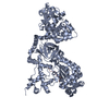
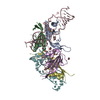
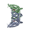
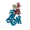
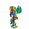
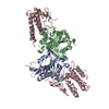
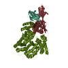
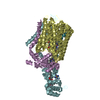
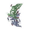
- PDB-3qnq: Crystal structure of the transporter ChbC, the IIC component from... -
+
Open data
ID or keywords:
Loading...
-
Basic information
Entry
Database: PDB / ID: 3qnq
Title
Crystal structure of the transporter ChbC, the IIC component from the N,N'-diacetylchitobiose-specific phosphotransferase system
Components
PTS system, cellobiose-specific IIC component
Keywords
membrane protein / transport protein / transporter / Enzyme IIC / phosphoenolpyruvate-sugar phosphotransferase system / Structural Genomics / PSI-Biology / New York Consortium on Membrane Protein Structure / NYCOMPS / hydrolase
Function / homology
Function and homology information
carbohydrate derivative transport / protein-N(PI)-phosphohistidine-carbohydrate phosphotransferase activity / phosphoenolpyruvate-dependent sugar phosphotransferase system / identical protein binding / plasma membrane Similarity search - Function
Redundancy: 10.1 % / Av σ(I) over netI: 26.64 / Number: 478665 / Rmerge(I) obs: 0.117 / Χ2: 0.96 / D res high: 4.5 Å / D res low: 50 Å / Num. obs: 24476 / % possible obs: 98.2
Diffraction reflection shell
Highest resolution (Å)
Lowest resolution (Å)
% possible obs (%)
ID
Rmerge(I) obs
Chi squared
Redundancy
9.67
50
99.6
1
0.071
0.999
12.9
7.69
9.67
100
1
0.089
1
10.9
6.72
7.69
100
1
0.2
1
9.6
6.11
6.72
100
1
0.372
0.963
9.3
5.67
6.11
100
1
0.559
0.926
9.4
5.33
5.67
100
1
0.586
0.904
9.5
5.07
5.33
100
1
0.494
0.898
9.6
4.85
5.07
99
1
0.32
0.961
9.7
4.66
4.85
94.9
1
0.297
0.966
9.7
4.5
4.66
87.6
1
0.379
0.953
9.8
Reflection
Resolution: 3.295→50 Å / Num. obs: 59563 / % possible obs: 95.7 % / Redundancy: 10.1 % / Rmerge(I) obs: 0.077 / Χ2: 0.871 / Net I/σ(I): 7.9
Reflection shell
Resolution (Å)
Redundancy (%)
Rmerge(I) obs
Num. unique all
Χ2
Diffraction-ID
% possible all
3.3-3.42
9.8
0.702
5569
0.697
1,2
91.4
3.42-3.55
9.7
0.443
5672
0.709
1,2
93.1
3.55-3.72
9.7
0.308
5644
0.74
1,2
92.5
3.72-3.91
9.6
0.208
5699
0.773
1,2
92.6
3.91-4.16
9.5
0.14
5797
0.83
1,2
94.5
4.16-4.48
9.4
0.093
5902
0.918
1,2
95.8
4.48-4.93
9.3
0.07
6078
0.946
1,2
97.7
4.93-5.64
9.6
0.075
6204
0.965
1,2
99.8
5.64-7.1
10.9
0.077
6337
1.011
1,2
100
7.1-50
12.9
0.042
6661
0.995
1,2
99.5
-
Phasing
Phasing
Method: SAD
-
Processing
Software
Name
Version
Classification
NB
DENZO
datareduction
SCALEPACK
datascaling
SHELX
phasing
DM
phasing
PHENIX
1.6.4_486
refinement
PDB_EXTRACT
3.1
dataextraction
HKL-2000
datacollection
HKL-2000
datareduction
HKL-2000
datascaling
SHARP
phasing
Refinement
Method to determine structure: SAD / Resolution: 3.295→19.974 Å / Occupancy max: 1 / Occupancy min: 1 / FOM work R set: 0.7701 / SU ML: 0.42 / σ(F): 1.34 / Stereochemistry target values: ML
Rfactor
Num. reflection
% reflection
Selection details
Rfree
0.2636
2996
5.08 %
RANDOM
Rwork
0.2275
-
-
-
obs
0.2294
58996
95.02 %
-
Solvent computation
Shrinkage radii: 0.95 Å / VDW probe radii: 1.2 Å / Solvent model: FLAT BULK SOLVENT MODEL / Bsol: 49.794 Å2 / ksol: 0.237 e/Å3
In the structure databanks used in Yorodumi, some data are registered as the other names, "COVID-19 virus" and "2019-nCoV". Here are the details of the virus and the list of structure data.
Jan 31, 2019. EMDB accession codes are about to change! (news from PDBe EMDB page)
EMDB accession codes are about to change! (news from PDBe EMDB page)
The allocation of 4 digits for EMDB accession codes will soon come to an end. Whilst these codes will remain in use, new EMDB accession codes will include an additional digit and will expand incrementally as the available range of codes is exhausted. The current 4-digit format prefixed with “EMD-” (i.e. EMD-XXXX) will advance to a 5-digit format (i.e. EMD-XXXXX), and so on. It is currently estimated that the 4-digit codes will be depleted around Spring 2019, at which point the 5-digit format will come into force.
The EM Navigator/Yorodumi systems omit the EMD- prefix.
Related info.:Q: What is EMD? / ID/Accession-code notation in Yorodumi/EM Navigator
Yorodumi is a browser for structure data from EMDB, PDB, SASBDB, etc.
This page is also the successor to EM Navigator detail page, and also detail information page/front-end page for Omokage search.
The word "yorodu" (or yorozu) is an old Japanese word meaning "ten thousand". "mi" (miru) is to see.
Related info.:EMDB / PDB / SASBDB / Comparison of 3 databanks / Yorodumi Search / Aug 31, 2016. New EM Navigator & Yorodumi / Yorodumi Papers / Jmol/JSmol / Function and homology information / Changes in new EM Navigator and Yorodumi
 Movie
Movie Controller

 Yorodumi
 Open data
 Basic information
 Components
 Keywords
 Function and homology information
 X-RAY DIFFRACTION /
  Authors
 Citation
 Structure visualization
 Downloads & links
 Other downloads
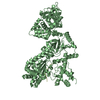

 PDBj
PDBj


 Assembly
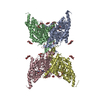
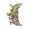


 Type: saccharide / Mass: 468.536 Da / Num. of mol.: 4
 Mass: 192.124 Da / Num. of mol.: 7 / Source method: obtained synthetically / Formula: C6H8O7
 Sample preparation
 Processing