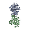
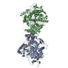
Entry Database : PDB / ID : 3bl8Title Crystal structure of the extracellular domain of neuroligin 2A from mouse Neuroligin-2 Keywords / / / / / Function / homology Function Domain/homology Component
/ / / / / / / / / / / / / / / / / / / / / / / / / / / / / / / / / / / / / / / / / / / / / / / / / / / / / / / / / / / / / / / / / / / / / / / / / / / Biological species Mus musculus (house mouse)Method / / / / Resolution : 3.3 Å Authors Jin, X. / Koehnke, J. / Shapiro, L. Journal : Proc.Natl.Acad.Sci.Usa / Year : 2008Title : Crystal structure of the extracellular cholinesterase-like domain from neuroligin-2.Authors : Koehnke, J. / Jin, X. / Budreck, E.C. / Posy, S. / Scheiffele, P. / Honig, B. / Shapiro, L. History Deposition Dec 10, 2007 Deposition site / Processing site Revision 1.0 Feb 19, 2008 Provider / Type Revision 1.1 Jul 13, 2011 Group / Version format complianceRevision 1.2 Oct 25, 2017 Group / Refinement description / Category / software / Item Revision 2.0 Jul 29, 2020 Group Advisory / Atomic model ... Advisory / Atomic model / Data collection / Database references / Derived calculations / Structure summary Category atom_site / chem_comp ... atom_site / chem_comp / database_PDB_caveat / entity / pdbx_branch_scheme / pdbx_chem_comp_identifier / pdbx_entity_branch / pdbx_entity_branch_descriptor / pdbx_entity_branch_link / pdbx_entity_branch_list / pdbx_entity_nonpoly / pdbx_nonpoly_scheme / pdbx_struct_assembly_gen / pdbx_unobs_or_zero_occ_atoms / pdbx_validate_chiral / pdbx_validate_close_contact / struct_asym / struct_conn / struct_ref_seq_dif / struct_site / struct_site_gen Item _atom_site.B_iso_or_equiv / _atom_site.Cartn_x ... _atom_site.B_iso_or_equiv / _atom_site.Cartn_x / _atom_site.Cartn_y / _atom_site.Cartn_z / _atom_site.auth_asym_id / _atom_site.auth_atom_id / _atom_site.auth_comp_id / _atom_site.auth_seq_id / _atom_site.label_asym_id / _atom_site.label_atom_id / _atom_site.label_comp_id / _atom_site.label_entity_id / _atom_site.type_symbol / _chem_comp.name / _chem_comp.type / _entity.formula_weight / _entity.pdbx_description / _entity.pdbx_number_of_molecules / _entity.src_method / _entity.type / _pdbx_struct_assembly_gen.asym_id_list / _pdbx_unobs_or_zero_occ_atoms.label_asym_id / _pdbx_validate_chiral.auth_asym_id / _pdbx_validate_chiral.auth_seq_id / _pdbx_validate_close_contact.auth_asym_id_2 / _pdbx_validate_close_contact.auth_seq_id_2 / _struct_conn.pdbx_leaving_atom_flag / _struct_conn.pdbx_role / _struct_conn.ptnr1_auth_asym_id / _struct_conn.ptnr1_auth_seq_id / _struct_conn.ptnr1_label_asym_id / _struct_conn.ptnr2_auth_asym_id / _struct_conn.ptnr2_auth_seq_id / _struct_conn.ptnr2_label_asym_id / _struct_ref_seq_dif.details Description / Provider / Type Revision 2.1 Mar 31, 2021 Group / Structure summary / Category / entity_src_genItem / _entity_src_gen.pdbx_host_org_cell_line / _entity_src_gen.pdbx_host_org_strainRevision 2.2 Oct 30, 2024 Group / Database references / Structure summaryCategory chem_comp_atom / chem_comp_bond ... chem_comp_atom / chem_comp_bond / database_2 / pdbx_entry_details / pdbx_modification_feature Item / _database_2.pdbx_database_accession
Show all Show less
 Movie
Movie Controller

 Yorodumi
 Open data
 Basic information
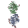
 Components
 Keywords
 Function and homology information
 X-RAY DIFFRACTION /
  Authors
 Citation
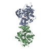
 Structure visualization
 Downloads & links
 Other downloads
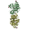


 PDBj
PDBj
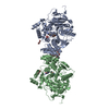
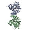
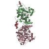
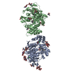
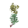
 Assembly
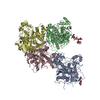
 Homo sapiens (human) / References: UniProt: Q69ZK9
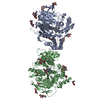
 Type: D-saccharide, beta linking / Mass: 221.208 Da / Num. of mol.: 2 / Source method: obtained synthetically / Formula: C8H15NO6
 Mass: 18.015 Da / Num. of mol.: 52 / Source method: isolated from a natural source / Formula: H2O
 Sample preparation
 / Beamline: X4C / Wavelength: 0.9793 Å
 Processing