 Movie
Movie Controller

[English] 日本語
 Yorodumi
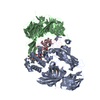
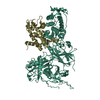
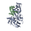
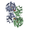
- PDB-1jmx: crystal structure of a quinohemoprotein amine dehydrogenase from ... -
+ Open data

- Basic information
| Entry | Database: PDB / ID: 1jmx | ||||||
|---|---|---|---|---|---|---|---|
| Title | crystal structure of a quinohemoprotein amine dehydrogenase from pseudomonas putida | ||||||
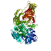
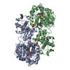
 Components Components | (Amine Dehydrogenase) x 3 | ||||||
 Keywords Keywords | OXIDOREDUCTASE / Amine Dehydrogenase | ||||||
| Function / homology |  Function and homology information Function and homology informationOxidoreductases; Acting on the CH-NH2 group of donors; With a copper protein as acceptor / aliphatic amine dehydrogenase activity / electron transfer activity / periplasmic space / heme binding / metal ion binding Similarity search - Function | ||||||
| Biological species |  Pseudomonas putida (bacteria) Pseudomonas putida (bacteria) | ||||||
| Method |  X-RAY DIFFRACTION / SYNCHROTRON / MIR / Resolution: 1.9 Å X-RAY DIFFRACTION / SYNCHROTRON / MIR / Resolution: 1.9 Å | ||||||
 Authors Authors | Satoh, A. / Miyahara, I. / Hirotsu, K. | ||||||
 Citation Citation | Journal: J.Biol.Chem. / Year: 2002 Title: Crystal structure of quinohemoprotein amine dehydrogenase from Pseudomonas putida. Identification of a novel quinone cofactor encaged by multiple thioether cross-bridges. Authors: Satoh, A. / Kim, J.K. / Miyahara, I. / Devreese, B. / Vandenberghe, I. / Hacisalihoglu, A. / Okajima, T. / Kuroda, S. / Adachi, O. / Duine, J.A. / Van Beeumen, J. / Tanizawa, K. / Hirotsu, K. | ||||||
| History |
|
- Structure visualization
| Structure viewer | Molecule: MolmilJmol/JSmol |
|---|
- Downloads & links
-Download
| PDBx/mmCIF format | 1jmx.cif.gz | 200 KB | Display | PDBx/mmCIF format |
|---|---|---|---|---|
| PDB format | pdb1jmx.ent.gz | 156.4 KB | Display | PDB format |
| PDBx/mmJSON format | 1jmx.json.gz | Tree view | PDBx/mmJSON format | |
| Others |  Other downloads Other downloads |
-Validation report
| Arichive directory | https://data.pdbj.org/pub/pdb/validation_reports/jm/1jmxftp://data.pdbj.org/pub/pdb/validation_reports/jm/1jmx | HTTPS FTP |
|---|
-Related structure data


-Links
 PDBj
PDBj


- Assembly
| Deposited unit | 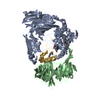
| ||||||||
|---|---|---|---|---|---|---|---|---|---|
| 1 |
| ||||||||
| 2 | 
| ||||||||
| Unit cell |
|
-Components
-Protein , 3 types, 3 molecules ABG
| #1: Protein | Mass: 53986.910 Da / Num. of mol.: 1 / Source method: isolated from a natural source / Source: (natural) Pseudomonas putida (bacteria) / References: UniProt: Q8VW85 |
|---|---|
| #2: Protein | Mass: 39284.551 Da / Num. of mol.: 1 / Source method: isolated from a natural source / Source: (natural) Pseudomonas putida (bacteria) / References: UniProt: Q8VW82 |
| #3: Protein | Mass: 8630.408 Da / Num. of mol.: 1 / Source method: isolated from a natural source / Source: (natural) Pseudomonas putida (bacteria) / References: UniProt: P0A182 |
-Non-polymers , 3 types, 460 molecules 


| #4: Chemical | ChemComp-NI /  Mass: 58.693 Da / Num. of mol.: 1 / Source method: obtained synthetically / Formula: Ni Mass: 58.693 Da / Num. of mol.: 1 / Source method: obtained synthetically / Formula: Ni | ||
|---|---|---|---|
| #5: Chemical |  Mass: 618.503 Da / Num. of mol.: 2 / Source method: obtained synthetically / Formula: C34H34FeN4O4 Mass: 618.503 Da / Num. of mol.: 2 / Source method: obtained synthetically / Formula: C34H34FeN4O4#6: Water | ChemComp-HOH / | Mass: 18.015 Da / Num. of mol.: 457 / Source method: isolated from a natural source / Formula: H2O |
-Details
| Has protein modification | Y |
|---|
-Experimental details
-Experiment
| Experiment | Method: X-RAY DIFFRACTION / Number of used crystals: 1 |
|---|
- Sample preparation
| Crystal | Density Matthews: 2.78 Å3/Da / Density % sol: 55.83 % | ||||||||||||||||||||||||
|---|---|---|---|---|---|---|---|---|---|---|---|---|---|---|---|---|---|---|---|---|---|---|---|---|---|
| Crystal grow | Temperature: 293 K / Method: vapor diffusion, hanging drop Details: PEG MME2000, nickel chloride, VAPOR DIFFUSION, HANGING DROP, temperature 293.0K | ||||||||||||||||||||||||
| Crystal grow | *PLUS Temperature: 20 ℃ | ||||||||||||||||||||||||
| Components of the solutions | *PLUS
|
-Data collection
| Diffraction | Mean temperature: 90 K |
|---|---|
| Diffraction source | Source: SYNCHROTRON / Site: Photon Factory  / Beamline: BL-18B / Wavelength: 1 Å / Beamline: BL-18B / Wavelength: 1 Å |
| Detector | Type: ADSC QUANTUM 4 / Detector: CCD / Date: Feb 20, 2001 |
| Radiation | Protocol: SINGLE WAVELENGTH / Monochromatic (M) / Laue (L): M / Scattering type: x-ray |
| Radiation wavelength | Wavelength: 1 Å / Relative weight: 1 |
| Reflection | Resolution: 1.9→20 Å / Num. obs: 89255 / % possible obs: 99.9 % / Observed criterion σ(F): 2 |
| Reflection | *PLUS Rmerge(I) obs: 0.051 |
| Reflection shell | *PLUS % possible obs: 100 % / Rmerge(I) obs: 0.26 / Mean I/σ(I) obs: 2.8 |
- Processing
| Software |
| ||||||||||||||||||
|---|---|---|---|---|---|---|---|---|---|---|---|---|---|---|---|---|---|---|---|
| Refinement | Method to determine structure: MIR / Resolution: 1.9→10 Å / σ(F): 2 / Stereochemistry target values: Engh & Huber
| ||||||||||||||||||
| Refinement step | Cycle: LAST / Resolution: 1.9→10 Å
| ||||||||||||||||||
| Refine LS restraints |
| ||||||||||||||||||
| Refinement | *PLUS σ(F): 2 / Rfactor obs: 0.211 / Rfactor Rfree: 0.245 | ||||||||||||||||||
| Solvent computation | *PLUS | ||||||||||||||||||
| Displacement parameters | *PLUS | ||||||||||||||||||
| LS refinement shell | *PLUS Rfactor Rfree: 0.296 / Rfactor obs: 0.237 |
