 Movie
Movie Controller


+ Open data

- Basic information
| Entry | Database: PDB / ID: 1df0 | ||||||
|---|---|---|---|---|---|---|---|
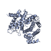
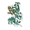
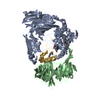
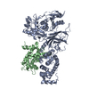
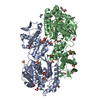
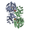
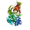
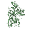
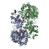
| Title | Crystal structure of M-Calpain | ||||||
 Components Components |
| ||||||
 Keywords Keywords | HYDROLASE / CYSTEINE PROTEASE / CALMODULIN / PAPAIN / CATALYTIC TRIAD / ZYMOGEN ACTIVATION / C2 DOMAIN / PROTEASE / ZYMOGEN / CALPAIN | ||||||
| Function / homology |  Function and homology information Function and homology informationcalpain-2 / Turbulent (oscillatory, disturbed) flow shear stress activates signaling by PIEZO1 and integrins in endothelial cells / Degradation of the extracellular matrix / calpain complex / positive regulation of phosphatidylcholine biosynthetic process / protein catabolic process at postsynapse / calcium-dependent cysteine-type endopeptidase activity / perinuclear endoplasmic reticulum / myoblast fusion / High laminar flow shear stress activates signaling by PIEZO1 and PECAM1:CDH5:KDR in endothelial cells ...calpain-2 / Turbulent (oscillatory, disturbed) flow shear stress activates signaling by PIEZO1 and integrins in endothelial cells / Degradation of the extracellular matrix / calpain complex / positive regulation of phosphatidylcholine biosynthetic process / protein catabolic process at postsynapse / calcium-dependent cysteine-type endopeptidase activity / perinuclear endoplasmic reticulum / myoblast fusion / High laminar flow shear stress activates signaling by PIEZO1 and PECAM1:CDH5:KDR in endothelial cells / regulation of interleukin-6 production / positive regulation of myoblast fusion / protein autoprocessing / pseudopodium / behavioral response to pain / blastocyst development / response to mechanical stimulus / synaptic vesicle endocytosis / cellular response to interferon-beta / cytoskeletal protein binding / cell projection / positive regulation of cardiac muscle cell apoptotic process / : / protein catabolic process / response to hydrogen peroxide / cellular response to amino acid stimulus / female pregnancy / peptidase activity / presynapse / cellular response to lipopolysaccharide / response to hypoxia / lysosome / postsynapse / membrane raft / external side of plasma membrane / focal adhesion / neuronal cell body / calcium ion binding / dendrite / chromatin / protein-containing complex binding / enzyme binding / Golgi apparatus / endoplasmic reticulum / proteolysis / membrane / nucleus / plasma membrane / cytoplasm / cytosol Similarity search - Function | ||||||
| Biological species |  | ||||||
| Method |  X-RAY DIFFRACTION / SYNCHROTRON / Resolution: 2.6 Å X-RAY DIFFRACTION / SYNCHROTRON / Resolution: 2.6 Å | ||||||
 Authors Authors | Hosfield, C.M. / Elce, J.S. / Davies, P.L. / Jia, Z. | ||||||
 Citation Citation | Journal: EMBO J. / Year: 1999 Title: Crystal structure of calpain reveals the structural basis for Ca(2+)-dependent protease activity and a novel mode of enzyme activation. Authors: Hosfield, C.M. / Elce, J.S. / Davies, P.L. / Jia, Z. #1: Journal: Nat.Struct.Biol. / Year: 1997Title: Structure of a Calpain Ca(2+)-Binding Domain Reveals a Novel EF-Hand and Ca(2+) -Induced Conformational Changes Authors: Blanchard, H. / Grochulski, P. / Li, Y. / Arthur, J.S.C. / Davies, P.L. / Elce, J.S. / Cygler, M. #2: Journal: Nat.Struct.Biol. / Year: 1997Title: Crystal Structure of Calcium Bound Domain VI of Calpain at 1.9 A Resolution and its Role in Enzyme Assembly, Regulation, and Inhibitor Binding Authors: Lin, G.D. / Chattopadhyay, D. / Maki, M. / Wang, K.K. / Carson, M. / Jin, L. / Hatanaka, M. / Takano, E. / Narayana, S.V. #3: Journal: Acta Crystallogr.,Sect.D / Year: 1999Title: Crystallization and X-Ray Crystallographic Analysis of M-Calpain: A Ca2+- Dependent Protease Authors: Hosfield, C.M. / Ye, Q. / Arthur, J.S.C. / Hegadorn, C. / Croall, D.E. / Elce, J.S. / Jia, Z. | ||||||
| History |
|
- Structure visualization
| Structure viewer | Molecule: MolmilJmol/JSmol |
|---|
- Downloads & links
-Download
| PDBx/mmCIF format | 1df0.cif.gz | 181.1 KB | Display | PDBx/mmCIF format |
|---|---|---|---|---|
| PDB format | pdb1df0.ent.gz | 142.8 KB | Display | PDB format |
| PDBx/mmJSON format | 1df0.json.gz | Tree view | PDBx/mmJSON format | |
| Others |  Other downloads Other downloads |
-Validation report
| Arichive directory | https://data.pdbj.org/pub/pdb/validation_reports/df/1df0ftp://data.pdbj.org/pub/pdb/validation_reports/df/1df0 | HTTPS FTP |
|---|
-Related structure data
| Related structure data | |
|---|---|
| Similar structure data |


-Links
 PDBj
PDBj


- Assembly
| Deposited unit | 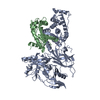
| ||||||||
|---|---|---|---|---|---|---|---|---|---|
| 1 |
| ||||||||
| Unit cell |
| ||||||||
| Details | The biological assembly is a heterodimer constructed from chain A and chain B |
-Components
| #1: Protein | Mass: 79994.039 Da / Num. of mol.: 1 / Fragment: LARGE (CATALYTIC) SUBUNIT / Mutation: C105S Source method: isolated from a genetically manipulated source Source: (gene. exp.)  |
|---|---|
| #2: Protein | Mass: 21304.979 Da / Num. of mol.: 1 Fragment: DOMAIN VI (CALCIUM-BINDING DOMAIN), SMALL (REGULATORY) SUBUNIT Source method: isolated from a genetically manipulated source Source: (gene. exp.) |
| #3: Water | ChemComp-HOH /  Mass: 18.015 Da / Num. of mol.: 358 / Source method: isolated from a natural source / Formula: H2O Mass: 18.015 Da / Num. of mol.: 358 / Source method: isolated from a natural source / Formula: H2O |
-Experimental details
-Experiment
| Experiment | Method: X-RAY DIFFRACTION / Number of used crystals: 1 |
|---|
- Sample preparation
| Crystal | Density Matthews: 3.03 Å3/Da / Density % sol: 59.34 % | |||||||||||||||||||||||||||||||||||
|---|---|---|---|---|---|---|---|---|---|---|---|---|---|---|---|---|---|---|---|---|---|---|---|---|---|---|---|---|---|---|---|---|---|---|---|---|
| Crystal grow | Temperature: 298 K / Method: vapor diffusion, hanging drop / pH: 6.25 Details: PEG 6000, MES, SODIUM CHLORIDE, DITHIOTHREITOL, EDTA, pH 6.25, VAPOR DIFFUSION, HANGING DROP | |||||||||||||||||||||||||||||||||||
| Crystal grow | *PLUS pH: 6.5 Details: Hosfield, C.M., (1999) Acta Crystallogr., Sect.D, 55, 1484. | |||||||||||||||||||||||||||||||||||
| Components of the solutions | *PLUS
|
-Data collection
| Diffraction | Mean temperature: 100 K |
|---|---|
| Diffraction source | Source: SYNCHROTRON / Site: CHESS  / Beamline: F2 / Wavelength: 0.923 / Beamline: F2 / Wavelength: 0.923 |
| Detector | Type: ADSC QUANTUM 4 / Detector: CCD / Date: Aug 25, 1998 |
| Radiation | Protocol: SINGLE WAVELENGTH / Monochromatic (M) / Laue (L): M / Scattering type: x-ray |
| Radiation wavelength | Wavelength: 0.923 Å / Relative weight: 1 |
| Reflection | Resolution: 2.6→50 Å / Num. all: 227921 / Num. obs: 227921 / % possible obs: 97.7 % / Observed criterion σ(F): 1 / Observed criterion σ(I): 1 / Redundancy: 3.1 % / Biso Wilson estimate: 68.052 Å2 / Rmerge(I) obs: 0.041 / Net I/σ(I): 17.1 |
| Reflection shell | Resolution: 2.6→2.69 Å / Redundancy: 2.91 % / Rmerge(I) obs: 0.11 / % possible all: 98.1 |
| Reflection | *PLUS |
- Processing
| Software |
| ||||||||||||||||||||||||||||||||||||||||||||||||||||||||||||
|---|---|---|---|---|---|---|---|---|---|---|---|---|---|---|---|---|---|---|---|---|---|---|---|---|---|---|---|---|---|---|---|---|---|---|---|---|---|---|---|---|---|---|---|---|---|---|---|---|---|---|---|---|---|---|---|---|---|---|---|---|---|
| Refinement | Resolution: 2.6→25 Å / σ(F): 0 / σ(I): 0 / Stereochemistry target values: ENGH & HUBER
| ||||||||||||||||||||||||||||||||||||||||||||||||||||||||||||
| Refinement step | Cycle: LAST / Resolution: 2.6→25 Å
| ||||||||||||||||||||||||||||||||||||||||||||||||||||||||||||
| Refine LS restraints |
| ||||||||||||||||||||||||||||||||||||||||||||||||||||||||||||
| Software | *PLUS Name: CNS / Classification: refinement | ||||||||||||||||||||||||||||||||||||||||||||||||||||||||||||
| Refinement | *PLUS Highest resolution: 2.6 Å / Lowest resolution: 25 Å / σ(F): 0 | ||||||||||||||||||||||||||||||||||||||||||||||||||||||||||||
| Solvent computation | *PLUS | ||||||||||||||||||||||||||||||||||||||||||||||||||||||||||||
| Displacement parameters | *PLUS | ||||||||||||||||||||||||||||||||||||||||||||||||||||||||||||
| Refine LS restraints | *PLUS
|
