 Movie
Movie Controller


+ Open data

- Basic information
| Entry | Database: PDB / ID: 1alw | ||||||
|---|---|---|---|---|---|---|---|
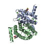
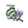
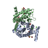
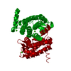
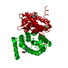
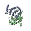
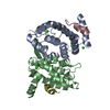
| Title | INHIBITOR AND CALCIUM BOUND DOMAIN VI OF PORCINE CALPAIN | ||||||
 Components Components | CALPAIN | ||||||
 Keywords Keywords | CALCIUM BINDING / CALMODULIN LIKE / DOMAIN OF CYSTEIN PROTEASE | ||||||
| Function / homology |  Function and homology information Function and homology informationHigh laminar flow shear stress activates signaling by PIEZO1 and PECAM1:CDH5:KDR in endothelial cells / Turbulent (oscillatory, disturbed) flow shear stress activates signaling by PIEZO1 and integrins in endothelial cells / Degradation of the extracellular matrix / calpain complex / calcium-dependent cysteine-type endopeptidase activity / calcium ion binding / proteolysis / plasma membrane / cytoplasm Similarity search - Function | ||||||
| Biological species |  | ||||||
| Method |  X-RAY DIFFRACTION / MOLECULAR REPLACEMENT / Resolution: 2.03 Å X-RAY DIFFRACTION / MOLECULAR REPLACEMENT / Resolution: 2.03 Å | ||||||
 Authors Authors | Narayana, S.V.L. / Lin, G. | ||||||
 Citation Citation | Journal: Nat.Struct.Biol. / Year: 1997 Title: Crystal structure of calcium bound domain VI of calpain at 1.9 A resolution and its role in enzyme assembly, regulation, and inhibitor binding. Authors: Lin, G.D. / Chattopadhyay, D. / Maki, M. / Wang, K.K. / Carson, M. / Jin, L. / Yuen, P.W. / Takano, E. / Hatanaka, M. / DeLucas, L.J. / Narayana, S.V. | ||||||
| History |
|
- Structure visualization
| Structure viewer | Molecule: MolmilJmol/JSmol |
|---|
- Downloads & links
-Download
| PDBx/mmCIF format | 1alw.cif.gz | 86.5 KB | Display | PDBx/mmCIF format |
|---|---|---|---|---|
| PDB format | pdb1alw.ent.gz | 64.7 KB | Display | PDB format |
| PDBx/mmJSON format | 1alw.json.gz | Tree view | PDBx/mmJSON format | |
| Others |  Other downloads Other downloads |
-Validation report
| Arichive directory | https://data.pdbj.org/pub/pdb/validation_reports/al/1alwftp://data.pdbj.org/pub/pdb/validation_reports/al/1alw | HTTPS FTP |
|---|
-Related structure data


-Links
 PDBj
PDBj- Assembly
| Deposited unit | 
| ||||||||
|---|---|---|---|---|---|---|---|---|---|
| 1 |
| ||||||||
| Unit cell |
| ||||||||
| Noncrystallographic symmetry (NCS) | NCS oper: (Code: given Matrix: (-0.3834, 0.923124, 0.029108), Vector: |
-Components
| #1: Protein | Mass: 19883.477 Da / Num. of mol.: 2 / Fragment: INHIBITOR-BOUND CALCIUM BINDING DOMAIN VI Source method: isolated from a genetically manipulated source Source: (gene. exp.)  #2: Chemical | ChemComp-CA /   Mass: 40.078 Da / Num. of mol.: 8 / Source method: obtained synthetically / Formula: Ca Mass: 40.078 Da / Num. of mol.: 8 / Source method: obtained synthetically / Formula: Ca#3: Chemical |   Mass: 308.136 Da / Num. of mol.: 2 / Source method: obtained synthetically / Formula: C9H9IO2S Mass: 308.136 Da / Num. of mol.: 2 / Source method: obtained synthetically / Formula: C9H9IO2S#4: Water | ChemComp-HOH / |  Mass: 18.015 Da / Num. of mol.: 104 / Source method: isolated from a natural source / Formula: H2O Mass: 18.015 Da / Num. of mol.: 104 / Source method: isolated from a natural source / Formula: H2O |
|---|
-Experimental details
-Experiment
| Experiment | Method: X-RAY DIFFRACTION / Number of used crystals: 2 |
|---|
- Sample preparation
| Crystal | Density Matthews: 2.48 Å3/Da / Density % sol: 50 % |
|---|---|
| Crystal grow | pH: 7.4 Details: PROTEIN WAS CRYSTALLIZED FROM 12% PEG6000 20 MM CACL2, 50 MM CACODYLATE BUFFER, PH7.4 |
| Crystal grow | *PLUS Method: vapor diffusion, hanging drop |
| Components of the solutions | *PLUS Common name: PEG6000 |
-Data collection
| Diffraction | Mean temperature: 280 K |
|---|---|
| Diffraction source | Source: ROTATING ANODE / Type: RIGAKU RUH2R / Wavelength: 1.5418 |
| Detector | Type: SIEMENS / Detector: AREA DETECTOR / Date: Feb 1, 1997 / Details: MIRRORS |
| Radiation | Monochromator: NI FILTER / Monochromatic (M) / Laue (L): M / Scattering type: x-ray |
| Radiation wavelength | Wavelength: 1.5418 Å / Relative weight: 1 |
| Reflection | Resolution: 2.03→20 Å / Num. obs: 24588 / % possible obs: 92.3 % / Observed criterion σ(I): 2 / Redundancy: 3 % / Biso Wilson estimate: 31 Å2 / Rmerge(I) obs: 0.06 / Rsym value: 0.08 / Net I/σ(I): 4.5 |
| Reflection shell | Resolution: 2.03→2.2 Å / Redundancy: 2 % / Rmerge(I) obs: 0.05 / Mean I/σ(I) obs: 3.5 / Rsym value: 0.26 / % possible all: 90 |
| Reflection shell | *PLUS % possible obs: 90 % |
- Processing
| Software |
| ||||||||||||||||||||||||||||||||||||||||||||||||||||||||||||
|---|---|---|---|---|---|---|---|---|---|---|---|---|---|---|---|---|---|---|---|---|---|---|---|---|---|---|---|---|---|---|---|---|---|---|---|---|---|---|---|---|---|---|---|---|---|---|---|---|---|---|---|---|---|---|---|---|---|---|---|---|---|
| Refinement | Method to determine structure: MOLECULAR REPLACEMENT Starting model: S-CAMLD Resolution: 2.03→8 Å / Data cutoff high absF: 100000 / Data cutoff low absF: 0.001 / Cross valid method: THROUGHOUT / σ(F): 2
| ||||||||||||||||||||||||||||||||||||||||||||||||||||||||||||
| Displacement parameters | Biso mean: 38 Å2 | ||||||||||||||||||||||||||||||||||||||||||||||||||||||||||||
| Refinement step | Cycle: LAST / Resolution: 2.03→8 Å
| ||||||||||||||||||||||||||||||||||||||||||||||||||||||||||||
| Refine LS restraints |
| ||||||||||||||||||||||||||||||||||||||||||||||||||||||||||||
| Xplor file |
| ||||||||||||||||||||||||||||||||||||||||||||||||||||||||||||
| Software | *PLUS Name: X-PLOR / Version: 3.5 / Classification: refinement | ||||||||||||||||||||||||||||||||||||||||||||||||||||||||||||
| Refinement | *PLUS | ||||||||||||||||||||||||||||||||||||||||||||||||||||||||||||
| Solvent computation | *PLUS | ||||||||||||||||||||||||||||||||||||||||||||||||||||||||||||
| Displacement parameters | *PLUS |
