 Movie
Movie Controller

[English] 日本語
 Yorodumi
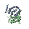
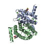
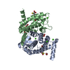
- PDB-4phj: The Structural Basis of Differential Inhibition of Human Calpain ... -
+ Open data

- Basic information
| Entry | Database: PDB / ID: 4phj | ||||||
|---|---|---|---|---|---|---|---|
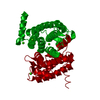
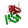
| Title | The Structural Basis of Differential Inhibition of Human Calpain by Indole and Phenyl alpha-Mercaptoacrylic Acids: Human unliganded protein | ||||||
 Components Components | Calpain small subunit 1 | ||||||
 Keywords Keywords | HYDROLASE / Domain VI / PEF(S) / Calcium Binding / Protease | ||||||
| Function / homology |  Function and homology information Function and homology informationcalpain complex / calcium-dependent cysteine-type endopeptidase activity / Formation of the cornified envelope / Deregulated CDK5 triggers multiple neurodegenerative pathways in Alzheimer's disease models / regulation of macroautophagy / Degradation of the extracellular matrix / Turbulent (oscillatory, disturbed) flow shear stress activates signaling by PIEZO1 and integrins in endothelial cells / High laminar flow shear stress activates signaling by PIEZO1 and PECAM1:CDH5:KDR in endothelial cells / calcium ion binding / positive regulation of cell population proliferation ...calpain complex / calcium-dependent cysteine-type endopeptidase activity / Formation of the cornified envelope / Deregulated CDK5 triggers multiple neurodegenerative pathways in Alzheimer's disease models / regulation of macroautophagy / Degradation of the extracellular matrix / Turbulent (oscillatory, disturbed) flow shear stress activates signaling by PIEZO1 and integrins in endothelial cells / High laminar flow shear stress activates signaling by PIEZO1 and PECAM1:CDH5:KDR in endothelial cells / calcium ion binding / positive regulation of cell population proliferation / proteolysis / extracellular exosome / membrane / plasma membrane / cytosol Similarity search - Function | ||||||
| Biological species |  Homo sapiens (human) Homo sapiens (human) | ||||||
| Method |  X-RAY DIFFRACTION / SYNCHROTRON / MOLECULAR REPLACEMENT / Resolution: 1.6 Å X-RAY DIFFRACTION / SYNCHROTRON / MOLECULAR REPLACEMENT / Resolution: 1.6 Å | ||||||
 Authors Authors | Adams, S.E. / Rizkallah, P.J. / Allemann, R.K. / Miller, D.J. / Hallett, M.B. / Robinson, E. | ||||||
 Citation Citation | Journal: J.Struct.Biol. / Year: 2014 Title: The structural basis of differential inhibition of human calpain by indole and phenyl alpha-mercaptoacrylic acids. Authors: Adams, S.E. / Rizkallah, P.J. / Miller, D.J. / Robinson, E.J. / Hallett, M.B. / Allemann, R.K. | ||||||
| History |
|
- Structure visualization
| Structure viewer | Molecule: MolmilJmol/JSmol |
|---|
- Downloads & links
-Download
| PDBx/mmCIF format | 4phj.cif.gz | 173.8 KB | Display | PDBx/mmCIF format |
|---|---|---|---|---|
| PDB format | pdb4phj.ent.gz | 135.9 KB | Display | PDB format |
| PDBx/mmJSON format | 4phj.json.gz | Tree view | PDBx/mmJSON format | |
| Others |  Other downloads Other downloads |
-Validation report
| Arichive directory | https://data.pdbj.org/pub/pdb/validation_reports/ph/4phjftp://data.pdbj.org/pub/pdb/validation_reports/ph/4phj | HTTPS FTP |
|---|
-Related structure data
| Related structure data |  4phkC 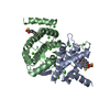 4phmC 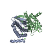 4phnC  1alvS S: Starting model for refinement C: citing same article ( |
|---|---|
| Similar structure data |


-Links
 PDBj
PDBj


- Assembly
| Deposited unit | 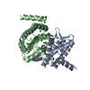
| |||||||||||||||||||||||||||
|---|---|---|---|---|---|---|---|---|---|---|---|---|---|---|---|---|---|---|---|---|---|---|---|---|---|---|---|---|
| 1 |
| |||||||||||||||||||||||||||
| Unit cell |
| |||||||||||||||||||||||||||
| Noncrystallographic symmetry (NCS) | NCS domain:
NCS domain segments:
|
-Components
| #1: Protein | Mass: 20016.607 Da / Num. of mol.: 2 / Fragment: Penta EF-hands subunit, Residues 96-268 Source method: isolated from a genetically manipulated source Source: (gene. exp.) Homo sapiens (human) / Gene: CAPNS1, CAPN4, CAPNS / Production host:  #2: Chemical | ChemComp-CA /   Mass: 40.078 Da / Num. of mol.: 8 / Source method: obtained synthetically / Formula: Ca Mass: 40.078 Da / Num. of mol.: 8 / Source method: obtained synthetically / Formula: Ca#3: Water | ChemComp-HOH / |  Mass: 18.015 Da / Num. of mol.: 376 / Source method: isolated from a natural source / Formula: H2O Mass: 18.015 Da / Num. of mol.: 376 / Source method: isolated from a natural source / Formula: H2O |
|---|
-Experimental details
-Experiment
| Experiment | Method: X-RAY DIFFRACTION |
|---|
- Sample preparation
| Crystal | Density Matthews: 2.81 Å3/Da / Density % sol: 56.17 % |
|---|---|
| Crystal grow | Temperature: 293 K / Method: vapor diffusion, sitting drop Details: PROTEIN WAS CRYSTALLIZED FROM 12% PEG6000, 20 MM CACL2, 50 MM CACODYLATE BUFFER, PH7.4 Temp details: Steady temperature |
-Data collection
| Diffraction | Mean temperature: 100 K |
|---|---|
| Diffraction source | Source: SYNCHROTRON / Site: Diamond  / Beamline: I04-1 / Wavelength: 0.92 Å / Beamline: I04-1 / Wavelength: 0.92 Å |
| Detector | Type: ADSC QUANTUM 315r / Detector: CCD / Date: Mar 16, 2013 |
| Radiation | Monochromator: Si (111) / Protocol: SINGLE WAVELENGTH / Monochromatic (M) / Laue (L): M / Scattering type: x-ray |
| Radiation wavelength | Wavelength: 0.92 Å / Relative weight: 1 |
| Reflection | Resolution: 1.6→49.61 Å / Num. all: 51828 / Num. obs: 51828 / % possible obs: 89 % / Redundancy: 4.1 % / Rmerge(I) obs: 0.05 / Net I/σ(I): 12.7 |
| Reflection shell | Resolution: 1.6→1.64 Å / Redundancy: 4.2 % / Rmerge(I) obs: 0.514 / Mean I/σ(I) obs: 2.3 / % possible all: 83.8 |
- Processing
| Software |
| ||||||||||||||||||||||||||||||||||||||||||||||||||||||||||||||||||||||||||||||||||||||||||||||||||||||||||||||||||||||||||||||||||||||||||||||||||||||||||||||||||||||||||||||||||||||
|---|---|---|---|---|---|---|---|---|---|---|---|---|---|---|---|---|---|---|---|---|---|---|---|---|---|---|---|---|---|---|---|---|---|---|---|---|---|---|---|---|---|---|---|---|---|---|---|---|---|---|---|---|---|---|---|---|---|---|---|---|---|---|---|---|---|---|---|---|---|---|---|---|---|---|---|---|---|---|---|---|---|---|---|---|---|---|---|---|---|---|---|---|---|---|---|---|---|---|---|---|---|---|---|---|---|---|---|---|---|---|---|---|---|---|---|---|---|---|---|---|---|---|---|---|---|---|---|---|---|---|---|---|---|---|---|---|---|---|---|---|---|---|---|---|---|---|---|---|---|---|---|---|---|---|---|---|---|---|---|---|---|---|---|---|---|---|---|---|---|---|---|---|---|---|---|---|---|---|---|---|---|---|---|
| Refinement | Method to determine structure: MOLECULAR REPLACEMENT Starting model: 1ALV Resolution: 1.6→49.61 Å / Cor.coef. Fo:Fc: 0.969 / Cor.coef. Fo:Fc free: 0.956 / Cross valid method: THROUGHOUT / ESU R: 0.088 / ESU R Free: 0.09 / Stereochemistry target values: MAXIMUM LIKELIHOOD / Details: HYDROGENS HAVE BEEN ADDED IN THE RIDING POSITIONS
| ||||||||||||||||||||||||||||||||||||||||||||||||||||||||||||||||||||||||||||||||||||||||||||||||||||||||||||||||||||||||||||||||||||||||||||||||||||||||||||||||||||||||||||||||||||||
| Solvent computation | Ion probe radii: 0.8 Å / Shrinkage radii: 0.8 Å / VDW probe radii: 1.2 Å / Solvent model: MASK | ||||||||||||||||||||||||||||||||||||||||||||||||||||||||||||||||||||||||||||||||||||||||||||||||||||||||||||||||||||||||||||||||||||||||||||||||||||||||||||||||||||||||||||||||||||||
| Displacement parameters | Biso mean: 25.583 Å2
| ||||||||||||||||||||||||||||||||||||||||||||||||||||||||||||||||||||||||||||||||||||||||||||||||||||||||||||||||||||||||||||||||||||||||||||||||||||||||||||||||||||||||||||||||||||||
| Refinement step | Cycle: 1 / Resolution: 1.6→49.61 Å
| ||||||||||||||||||||||||||||||||||||||||||||||||||||||||||||||||||||||||||||||||||||||||||||||||||||||||||||||||||||||||||||||||||||||||||||||||||||||||||||||||||||||||||||||||||||||
| Refine LS restraints |
|