 Movie
Movie Controller

[English] 日本語
 Yorodumi
- PDB-5jqp: Crystal structure of ER glucosidase II heterodimeric complex cons... -
+ Open data

- Basic information
| Entry | Database: PDB / ID: 5jqp | ||||||||||||||||||
|---|---|---|---|---|---|---|---|---|---|---|---|---|---|---|---|---|---|---|---|
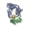
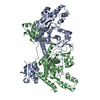
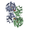
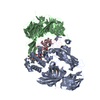
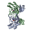
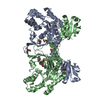
| Title | Crystal structure of ER glucosidase II heterodimeric complex consisting of catalytic subunit and the binding domain of regulatory subunit | ||||||||||||||||||
 Components Components |
| ||||||||||||||||||
 Keywords Keywords | HYDROLASE / PROTEIN TRANSPORT | ||||||||||||||||||
| Function / homology |  Function and homology information Function and homology informationglucosidase II complex / alpha-glucosidase / alpha-1,4-glucosidase activity / N-glycan processing / carbohydrate binding / carbohydrate metabolic process / metal ion binding Similarity search - Function | ||||||||||||||||||
| Biological species |  Chaetomium thermophilum (fungus) Chaetomium thermophilum (fungus) | ||||||||||||||||||
| Method |  X-RAY DIFFRACTION / SYNCHROTRON / MOLECULAR REPLACEMENT / Resolution: 2.2 Å X-RAY DIFFRACTION / SYNCHROTRON / MOLECULAR REPLACEMENT / Resolution: 2.2 Å | ||||||||||||||||||
 Authors Authors | Satoh, T. / Toshimori, T. / Noda, M. / Uchiyama, S. / Kato, K. | ||||||||||||||||||
| Funding support |  Japan, 5items Japan, 5items
| ||||||||||||||||||
 Citation Citation | Journal: Protein Sci. / Year: 2016 Title: Interaction mode between catalytic and regulatory subunits in glucosidase II involved in ER glycoprotein quality control. Authors: Satoh, T. / Toshimori, T. / Noda, M. / Uchiyama, S. / Kato, K. | ||||||||||||||||||
| History |
|
- Structure visualization
| Structure viewer | Molecule: MolmilJmol/JSmol |
|---|
- Downloads & links
-Download
| PDBx/mmCIF format | 5jqp.cif.gz | 241.1 KB | Display | PDBx/mmCIF format |
|---|---|---|---|---|
| PDB format | pdb5jqp.ent.gz | 185.1 KB | Display | PDB format |
| PDBx/mmJSON format | 5jqp.json.gz | Tree view | PDBx/mmJSON format | |
| Others |  Other downloads Other downloads |
-Validation report
| Arichive directory | https://data.pdbj.org/pub/pdb/validation_reports/jq/5jqpftp://data.pdbj.org/pub/pdb/validation_reports/jq/5jqp | HTTPS FTP |
|---|
-Related structure data
| Related structure data |  5dkxS S: Starting model for refinement |
|---|---|
| Similar structure data |


-Links
 PDBj
PDBj


- Assembly
| Deposited unit | 
| |||||||||
|---|---|---|---|---|---|---|---|---|---|---|
| 1 |
| |||||||||
| Unit cell |
| |||||||||
| Components on special symmetry positions |
|
-Components
| #1: Protein | Mass: 108501.156 Da / Num. of mol.: 1 Source method: isolated from a genetically manipulated source Source: (gene. exp.) Chaetomium thermophilum (strain DSM 1495 / CBS 144.50 / IMI 039719) (fungus)Strain: DSM 1495 / CBS 144.50 / IMI 039719 / Gene: CTHT_0064960 / Plasmid: PCOLD-GST (MODIFIED) / Production host:  | ||||
|---|---|---|---|---|---|
| #2: Protein | Mass: 17753.391 Da / Num. of mol.: 1 / Fragment: UNP RESIDUES 21-162 Source method: isolated from a genetically manipulated source Source: (gene. exp.) Chaetomium thermophilum (strain DSM 1495 / CBS 144.50 / IMI 039719) (fungus)Strain: DSM 1495 / CBS 144.50 / IMI 039719 / Gene: CTHT_0046400 / Plasmid: pET16b / Production host: | ||||
| #3: Chemical | ChemComp-TRS /   Mass: 122.143 Da / Num. of mol.: 1 / Source method: obtained synthetically / Formula: C4H12NO3 / Comment: pH buffer*YM Mass: 122.143 Da / Num. of mol.: 1 / Source method: obtained synthetically / Formula: C4H12NO3 / Comment: pH buffer*YM | ||||
| #4: Chemical |   Mass: 40.078 Da / Num. of mol.: 2 / Source method: obtained synthetically / Formula: Ca Mass: 40.078 Da / Num. of mol.: 2 / Source method: obtained synthetically / Formula: Ca#5: Water | ChemComp-HOH / |  Mass: 18.015 Da / Num. of mol.: 655 / Source method: isolated from a natural source / Formula: H2O Mass: 18.015 Da / Num. of mol.: 655 / Source method: isolated from a natural source / Formula: H2OHas protein modification | Y | |
-Experimental details
-Experiment
| Experiment | Method: X-RAY DIFFRACTION / Number of used crystals: 1 |
|---|
- Sample preparation
| Crystal | Density Matthews: 2.52 Å3/Da / Density % sol: 51.1 % |
|---|---|
| Crystal grow | Temperature: 293 K / Method: vapor diffusion, hanging drop / pH: 6.5 Details: 20% PEG 3350, 100 mM Bis-Tris (pH 6.5), 0.2 M ammonium phosphate |
-Data collection
| Diffraction | Mean temperature: 95 K |
|---|---|
| Diffraction source | Source: SYNCHROTRON / Site: Photon Factory / Beamline: BL-1A / Wavelength: 1.1 Å |
| Detector | Type: DECTRIS EIGER R 4M / Detector: PIXEL / Date: Dec 15, 2015 |
| Radiation | Protocol: SINGLE WAVELENGTH / Monochromatic (M) / Laue (L): M / Scattering type: x-ray |
| Radiation wavelength | Wavelength: 1.1 Å / Relative weight: 1 |
| Reflection | Resolution: 2.2→50 Å / Num. obs: 64671 / % possible obs: 99.4 % / Observed criterion σ(I): -3 / Redundancy: 4.4 % / Rmerge(I) obs: 0.108 / Net I/σ(I): 11.8 |
| Reflection shell | Resolution: 2.2→2.24 Å / Redundancy: 4.6 % / Rmerge(I) obs: 0.499 / Mean I/σ(I) obs: 2.2 / % possible all: 100 |
- Processing
| Software |
| ||||||||||||||||||||||||||||||||||||||||||||||||||||||||||||||||||||||||||||||||||||||||||||||||||||||||||||||||||||||||||||||||||||||||||||||||||||||||||||||||||||||||||||||||||||||
|---|---|---|---|---|---|---|---|---|---|---|---|---|---|---|---|---|---|---|---|---|---|---|---|---|---|---|---|---|---|---|---|---|---|---|---|---|---|---|---|---|---|---|---|---|---|---|---|---|---|---|---|---|---|---|---|---|---|---|---|---|---|---|---|---|---|---|---|---|---|---|---|---|---|---|---|---|---|---|---|---|---|---|---|---|---|---|---|---|---|---|---|---|---|---|---|---|---|---|---|---|---|---|---|---|---|---|---|---|---|---|---|---|---|---|---|---|---|---|---|---|---|---|---|---|---|---|---|---|---|---|---|---|---|---|---|---|---|---|---|---|---|---|---|---|---|---|---|---|---|---|---|---|---|---|---|---|---|---|---|---|---|---|---|---|---|---|---|---|---|---|---|---|---|---|---|---|---|---|---|---|---|---|---|
| Refinement | Method to determine structure: MOLECULAR REPLACEMENT Starting model: 5DKX Resolution: 2.2→20 Å / Cor.coef. Fo:Fc: 0.964 / Cor.coef. Fo:Fc free: 0.935 / SU B: 5.189 / SU ML: 0.127 / Cross valid method: THROUGHOUT / ESU R: 0.206 / ESU R Free: 0.178 / Details: HYDROGENS HAVE BEEN ADDED IN THE RIDING POSITIONS
| ||||||||||||||||||||||||||||||||||||||||||||||||||||||||||||||||||||||||||||||||||||||||||||||||||||||||||||||||||||||||||||||||||||||||||||||||||||||||||||||||||||||||||||||||||||||
| Solvent computation | Ion probe radii: 0.8 Å / Shrinkage radii: 0.8 Å / VDW probe radii: 1.2 Å | ||||||||||||||||||||||||||||||||||||||||||||||||||||||||||||||||||||||||||||||||||||||||||||||||||||||||||||||||||||||||||||||||||||||||||||||||||||||||||||||||||||||||||||||||||||||
| Displacement parameters | Biso mean: 25.227 Å2
| ||||||||||||||||||||||||||||||||||||||||||||||||||||||||||||||||||||||||||||||||||||||||||||||||||||||||||||||||||||||||||||||||||||||||||||||||||||||||||||||||||||||||||||||||||||||
| Refinement step | Cycle: 1 / Resolution: 2.2→20 Å
| ||||||||||||||||||||||||||||||||||||||||||||||||||||||||||||||||||||||||||||||||||||||||||||||||||||||||||||||||||||||||||||||||||||||||||||||||||||||||||||||||||||||||||||||||||||||
| Refine LS restraints |
|