 Movie
Movie Controller

[English] 日本語
 Yorodumi
- PDB-1pby: Structure of the Phenylhydrazine Adduct of the Quinohemoprotein A... -
+ Open data

- Basic information
| Entry | Database: PDB / ID: 1pby | |||||||||
|---|---|---|---|---|---|---|---|---|---|---|
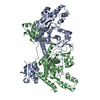
| Title | Structure of the Phenylhydrazine Adduct of the Quinohemoprotein Amine Dehydrogenase from Paracoccus denitrificans at 1.7 A Resolution | |||||||||
 Components Components | (quinohemoprotein amine dehydrogenase ...) x 3 | |||||||||
 Keywords Keywords | OXIDOREDUCTASE / QUINOHEMOPROTEIN / AMINE DEHYDROGENASE | |||||||||
| Function / homology |  Function and homology information Function and homology informationOxidoreductases; Acting on the CH-NH2 group of donors; With a cytochrome as acceptor / oxidoreductase activity, acting on the CH-NH2 group of donors / periplasmic space / electron transfer activity / heme binding / metal ion binding Similarity search - Function | |||||||||
| Biological species |  Paracoccus denitrificans (bacteria) Paracoccus denitrificans (bacteria) | |||||||||
| Method |  X-RAY DIFFRACTION / SYNCHROTRON / refined directly / Resolution: 1.7 Å X-RAY DIFFRACTION / SYNCHROTRON / refined directly / Resolution: 1.7 Å | |||||||||
 Authors Authors | Datta, S. / Ikeda, T. / Kano, K. / Mathews, F.S. | |||||||||
 Citation Citation | Journal: Acta Crystallogr.,Sect.D / Year: 2003 Title: Structure of the phenylhydrazine adduct of the quinohemoprotein amine dehydrogenase from Paracoccus denitrificans at 1.7 A resolution. Authors: Datta, S. / Ikeda, T. / Kano, K. / Mathews, F.S. #1: Journal: Proc.Natl.Acad.Sci.USA / Year: 2001Title: Structure of a quinohemoprotein amine dehydrogenase with an uncommon redox cofactor and highly unusual crosslinking | |||||||||
| History |
|
- Structure visualization
| Structure viewer | Molecule: MolmilJmol/JSmol |
|---|
- Downloads & links
-Download
| PDBx/mmCIF format | 1pby.cif.gz | 223.7 KB | Display | PDBx/mmCIF format |
|---|---|---|---|---|
| PDB format | pdb1pby.ent.gz | 172.1 KB | Display | PDB format |
| PDBx/mmJSON format | 1pby.json.gz | Tree view | PDBx/mmJSON format | |
| Others |  Other downloads Other downloads |
-Validation report
| Arichive directory | https://data.pdbj.org/pub/pdb/validation_reports/pb/1pbyftp://data.pdbj.org/pub/pdb/validation_reports/pb/1pby | HTTPS FTP |
|---|
-Related structure data
| Related structure data |  1jjuS S: Starting model for refinement |
|---|---|
| Similar structure data |


-Links
 PDBj
PDBj


- Assembly
| Deposited unit | 
| ||||||||
|---|---|---|---|---|---|---|---|---|---|
| 1 |
| ||||||||
| Unit cell |
|
-Components
-Quinohemoprotein amine dehydrogenase ... , 3 types, 3 molecules ABC
| #1: Protein | Mass: 52711.500 Da / Num. of mol.: 1 Source method: isolated from a genetically manipulated source Source: (gene. exp.) Paracoccus denitrificans (bacteria) / Production host: Paracoccus denitrificans (bacteria) / Strain (production host): IFO12442 / References: UniProt: Q8VUT0, EC: 1.4.99.3 |
|---|---|
| #2: Protein | Mass: 37021.953 Da / Num. of mol.: 1 Source method: isolated from a genetically manipulated source Source: (gene. exp.) Paracoccus denitrificans (bacteria) / Production host: Paracoccus denitrificans (bacteria) / References: GenBank: 17402571, UniProt: Q8VUS7*PLUS |
| #3: Protein | Mass: 8843.791 Da / Num. of mol.: 1 Source method: isolated from a genetically manipulated source Source: (gene. exp.) Paracoccus denitrificans (bacteria) / Production host: Paracoccus denitrificans (bacteria) / References: GenBank: 17402570, UniProt: Q8VUS8*PLUS |
-Non-polymers , 3 types, 1301 molecules 

| #4: Chemical |  Mass: 618.503 Da / Num. of mol.: 2 / Source method: obtained synthetically / Formula: C34H34FeN4O4 Mass: 618.503 Da / Num. of mol.: 2 / Source method: obtained synthetically / Formula: C34H34FeN4O4#5: Chemical | ChemComp-TBU /  Mass: 74.122 Da / Num. of mol.: 4 / Source method: obtained synthetically / Formula: C4H10O Mass: 74.122 Da / Num. of mol.: 4 / Source method: obtained synthetically / Formula: C4H10O#6: Water | ChemComp-HOH / | Mass: 18.015 Da / Num. of mol.: 1295 / Source method: isolated from a natural source / Formula: H2O |
|---|
-Experimental details
-Experiment
| Experiment | Method: X-RAY DIFFRACTION / Number of used crystals: 1 |
|---|
- Sample preparation
| Crystal | Density Matthews: 2.66 Å3/Da / Density % sol: 53.74 % | ||||||||||||||||||||||||
|---|---|---|---|---|---|---|---|---|---|---|---|---|---|---|---|---|---|---|---|---|---|---|---|---|---|
| Crystal grow | Temperature: 293 K / Method: vapor diffusion, sitting drop / pH: 5.6 Details: PEG 4000, sodium citrate, t-butyl alcohol, pH 5.6, VAPOR DIFFUSION, SITTING DROP, temperature 293K | ||||||||||||||||||||||||
| Crystal grow | *PLUS Method: vapor diffusion | ||||||||||||||||||||||||
| Components of the solutions | *PLUS
|
-Data collection
| Diffraction | Mean temperature: 100 K |
|---|---|
| Diffraction source | Source: SYNCHROTRON / Site: APS  / Beamline: 14-BM-C / Wavelength: 0.9 Å / Beamline: 14-BM-C / Wavelength: 0.9 Å |
| Detector | Type: ADSC QUANTUM 4 / Detector: CCD / Date: Dec 16, 2001 |
| Radiation | Monochromator: Si 111 / Protocol: SINGLE WAVELENGTH / Monochromatic (M) / Laue (L): M / Scattering type: x-ray |
| Radiation wavelength | Wavelength: 0.9 Å / Relative weight: 1 |
| Reflection | Resolution: 1.7→36.24 Å / Num. all: 117161 / Num. obs: 110131 / % possible obs: 94.2 % / Observed criterion σ(I): -3 / Redundancy: 6.76 % / Biso Wilson estimate: 17.2 Å2 / Rmerge(I) obs: 0.06 / Net I/σ(I): 27.7 |
| Reflection shell | Resolution: 1.7→1.76 Å / Redundancy: 1.3 % / Rmerge(I) obs: 0.481 / Mean I/σ(I) obs: 2.3 / Num. unique all: 8700 / % possible all: 75.4 |
| Reflection | *PLUS Highest resolution: 1.7 Å / Lowest resolution: 30 Å / Redundancy: 6.7 % / Rmerge(I) obs: 0.06 |
| Reflection shell | *PLUS Highest resolution: 1.7 Å / % possible obs: 75.4 % / Mean I/σ(I) obs: 2.4 |
- Processing
| Software |
| |||||||||||||||||||||||||||||||||||||||||||||||||||||||||||||||
|---|---|---|---|---|---|---|---|---|---|---|---|---|---|---|---|---|---|---|---|---|---|---|---|---|---|---|---|---|---|---|---|---|---|---|---|---|---|---|---|---|---|---|---|---|---|---|---|---|---|---|---|---|---|---|---|---|---|---|---|---|---|---|---|---|
| Refinement | Method to determine structure: refined directly Starting model: PDB ENTRY 1JJU Resolution: 1.7→36.42 Å / Rfactor Rfree error: 0.002 / Isotropic thermal model: Isotropic / Cross valid method: THROUGHOUT / σ(F): 0 / Stereochemistry target values: Engh & Huber
| |||||||||||||||||||||||||||||||||||||||||||||||||||||||||||||||
| Displacement parameters | Biso mean: 22.5 Å2 | |||||||||||||||||||||||||||||||||||||||||||||||||||||||||||||||
| Refine analyze |
| |||||||||||||||||||||||||||||||||||||||||||||||||||||||||||||||
| Refinement step | Cycle: LAST / Resolution: 1.7→36.42 Å
| |||||||||||||||||||||||||||||||||||||||||||||||||||||||||||||||
| Refine LS restraints |
| |||||||||||||||||||||||||||||||||||||||||||||||||||||||||||||||
| LS refinement shell | Refine-ID: X-RAY DIFFRACTION / Total num. of bins used: 6
| |||||||||||||||||||||||||||||||||||||||||||||||||||||||||||||||
| Refinement | *PLUS Lowest resolution: 30 Å / % reflection Rfree: 10 % | |||||||||||||||||||||||||||||||||||||||||||||||||||||||||||||||
| Solvent computation | *PLUS | |||||||||||||||||||||||||||||||||||||||||||||||||||||||||||||||
| Displacement parameters | *PLUS | |||||||||||||||||||||||||||||||||||||||||||||||||||||||||||||||
| Refine LS restraints | *PLUS
|
