 Movie
Movie Controller

[English] 日本語
 Yorodumi
- PDB-2mys: MYOSIN SUBFRAGMENT-1, ALPHA CARBON COORDINATES ONLY FOR THE TWO L... -
+ Open data

- Basic information
| Entry | Database: PDB / ID: 2mys | |||||||||
|---|---|---|---|---|---|---|---|---|---|---|
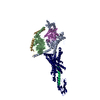
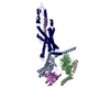
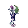
| Title | MYOSIN SUBFRAGMENT-1, ALPHA CARBON COORDINATES ONLY FOR THE TWO LIGHT CHAINS | |||||||||
 Components Components | (MYOSIN) x 3 | |||||||||
 Keywords Keywords | MUSCLE PROTEIN / MYOSIN SUBFRAGMENT-1 / MYOSIN HEAD / MOTOR PROTEIN | |||||||||
| Function / homology |  Function and homology information Function and homology informationregulation of myosin II filament assembly / contractile muscle fiber / Striated Muscle Contraction / myosin filament / myosin complex / myosin II complex / structural constituent of muscle / microfilament motor activity / myofibril / skeletal muscle tissue development ...regulation of myosin II filament assembly / contractile muscle fiber / Striated Muscle Contraction / myosin filament / myosin complex / myosin II complex / structural constituent of muscle / microfilament motor activity / myofibril / skeletal muscle tissue development / muscle contraction / actin filament binding / calmodulin binding / calcium ion binding / ATP binding / cytoplasm Similarity search - Function | |||||||||
| Biological species |  | |||||||||
| Method |  X-RAY DIFFRACTION / Resolution: 2.8 Å X-RAY DIFFRACTION / Resolution: 2.8 Å | |||||||||
 Authors Authors | Rayment, I. / Holden, H.M. | |||||||||
 Citation Citation | Journal: Science / Year: 1993 Title: Three-dimensional structure of myosin subfragment-1: a molecular motor. Authors: Rayment, I. / Rypniewski, W.R. / Schmidt-Base, K. / Smith, R. / Tomchick, D.R. / Benning, M.M. / Winkelmann, D.A. / Wesenberg, G. / Holden, H.M. #1: Journal: Biochemistry / Year: 1995Title: X-Ray Structures of the Myosin Motor Domain of Dictyostelium Discoideum Complexed with Mgadp.Befx and Mgadp.Alf4- Authors: Fisher, A.J. / Smith, C.A. / Thoden, J.B. / Smith, R. / Sutoh, K. / Holden, H.M. / Rayment, I. #2: Journal: Biochemistry / Year: 1995Title: X-Ray Structure of the Magnesium(II)-Pyrophosphate Complex of the Truncated Head of Dictyostelium Discoideum Myosin to 2.7 A Resolution Authors: Smith, C.A. / Rayment, I. | |||||||||
| History |
|
- Structure visualization
| Structure viewer | Molecule: MolmilJmol/JSmol |
|---|
- Downloads & links
-Download
| PDBx/mmCIF format | 2mys.cif.gz | 184.8 KB | Display | PDBx/mmCIF format |
|---|---|---|---|---|
| PDB format | pdb2mys.ent.gz | 145.3 KB | Display | PDB format |
| PDBx/mmJSON format | 2mys.json.gz | Tree view | PDBx/mmJSON format | |
| Others |  Other downloads Other downloads |
-Validation report
| Arichive directory | https://data.pdbj.org/pub/pdb/validation_reports/my/2mysftp://data.pdbj.org/pub/pdb/validation_reports/my/2mys | HTTPS FTP |
|---|
-Related structure data
| Similar structure data |
|---|


-Links
 PDBj
PDBj


- Assembly
| Deposited unit | 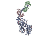
| ||||||||
|---|---|---|---|---|---|---|---|---|---|
| 1 |
| ||||||||
| Unit cell |
|
-Components
| #1: Protein | Mass: 96880.750 Da / Num. of mol.: 1 / Fragment: SUBFRAGMENT-1 / Source method: isolated from a natural source Details: PROTEOLYTIC FRAGMENT OF MYOSIN GENERATED BY PAPAIN DIGESTION Source: (natural) |
|---|---|
| #2: Protein | Mass: 18329.598 Da / Num. of mol.: 1 / Fragment: SUBFRAGMENT-1 / Source method: isolated from a natural source Details: PROTEOLYTIC FRAGMENT OF MYOSIN GENERATED BY PAPAIN DIGESTION Source: (natural) |
| #3: Protein | Mass: 16340.213 Da / Num. of mol.: 1 / Fragment: SUBFRAGMENT-1 / Source method: isolated from a natural source Details: PROTEOLYTIC FRAGMENT OF MYOSIN GENERATED BY PAPAIN DIGESTION Source: (natural) |
| #4: Chemical | ChemComp-SO4 /   Mass: 96.063 Da / Num. of mol.: 1 / Source method: obtained synthetically / Formula: SO4 Mass: 96.063 Da / Num. of mol.: 1 / Source method: obtained synthetically / Formula: SO4 |
| #5: Chemical | ChemComp-MG /   Mass: 24.305 Da / Num. of mol.: 1 / Source method: obtained synthetically / Formula: Mg Mass: 24.305 Da / Num. of mol.: 1 / Source method: obtained synthetically / Formula: Mg |
-Experimental details
-Experiment
| Experiment | Method: X-RAY DIFFRACTION |
|---|
- Sample preparation
| Crystal | Density Matthews: 3.19 Å3/Da / Density % sol: 61.46 % | |||||||||||||||||||||||||||||||||||
|---|---|---|---|---|---|---|---|---|---|---|---|---|---|---|---|---|---|---|---|---|---|---|---|---|---|---|---|---|---|---|---|---|---|---|---|---|
| Crystal grow | *PLUS pH: 6.7 / Method: batch method / Details: macroseeding | |||||||||||||||||||||||||||||||||||
| Components of the solutions | *PLUS
|
-Data collection
| Diffraction source | Wavelength: 1.5418 |
|---|---|
| Detector | Type: SIEMENS / Detector: AREA DETECTOR / Date: 1991 |
| Radiation | Monochromatic (M) / Laue (L): M / Scattering type: x-ray |
| Radiation wavelength | Wavelength: 1.5418 Å / Relative weight: 1 |
| Reflection | Redundancy: 3.8 % / Rmerge(I) obs: 0.053 |
| Reflection | *PLUS Highest resolution: 2.8 Å / Lowest resolution: 4.5 Å / Num. obs: 36781 / Num. measured all: 178986 / Rmerge(I) obs: 0.092 |
- Processing
| Software |
| |||||||||||||||
|---|---|---|---|---|---|---|---|---|---|---|---|---|---|---|---|---|
| Refinement | Resolution: 2.8→30 Å / σ(F): 0 /
| |||||||||||||||
| Refinement step | Cycle: LAST / Resolution: 2.8→30 Å
| |||||||||||||||
| Software | *PLUS Name: TNT / Classification: refinement | |||||||||||||||
| Refinement | *PLUS Rfactor all: 0.233 | |||||||||||||||
| Solvent computation | *PLUS | |||||||||||||||
| Displacement parameters | *PLUS | |||||||||||||||
| Refine LS restraints | *PLUS
|
