[English] 日本語
 Yorodumi
- PDB-2bbm: SOLUTION STRUCTURE OF A CALMODULIN-TARGET PEPTIDE COMPLEX BY MULT... -
+ Open data
- Basic information
| Entry | Database: PDB / ID: 2bbm | ||||||
|---|---|---|---|---|---|---|---|
| Title | SOLUTION STRUCTURE OF A CALMODULIN-TARGET PEPTIDE COMPLEX BY MULTIDIMENSIONAL NMR | ||||||
 Components Components |
| ||||||
 Keywords Keywords | CALCIUM-BINDING PROTEIN | ||||||
| Function / homology |  Function and homology information Function and homology informationnegative regulation of phospholipase C-activating phototransduction signaling pathway / myosin VI complex / myosin VI head/neck binding / myosin VII complex / photoreceptor cell axon guidance / negative regulation of opsin-mediated signaling pathway / rhabdomere development / regulation of muscle filament sliding / rhabdomere / myosin-light-chain kinase ...negative regulation of phospholipase C-activating phototransduction signaling pathway / myosin VI complex / myosin VI head/neck binding / myosin VII complex / photoreceptor cell axon guidance / negative regulation of opsin-mediated signaling pathway / rhabdomere development / regulation of muscle filament sliding / rhabdomere / myosin-light-chain kinase / myosin V complex / myosin light chain kinase activity / detection of chemical stimulus involved in sensory perception of smell / kinetochore organization / autophagic cell death / G protein-coupled opsin signaling pathway / actin filament-based movement / myosin V binding / channel regulator activity / calcium/calmodulin-dependent protein kinase activity / myosin heavy chain binding / muscle cell cellular homeostasis / mitotic spindle pole / centriole replication / cellular response to ethanol / enzyme regulator activity / centriole / sensory perception of sound / microtubule cytoskeleton organization / spindle / mitotic spindle / sensory perception of smell / midbody / cell cortex / calmodulin binding / calcium ion binding / centrosome / nucleoplasm / ATP binding / cytoplasm / cytosol Similarity search - Function | ||||||
| Biological species |  | ||||||
| Method | SOLUTION NMR | ||||||
 Authors Authors | Clore, G.M. / Bax, A. / Ikura, M. / Gronenborn, A.M. | ||||||
 Citation Citation |  Journal: Science / Year: 1992 Journal: Science / Year: 1992Title: Solution structure of a calmodulin-target peptide complex by multidimensional NMR. Authors: Ikura, M. / Clore, G.M. / Gronenborn, A.M. / Zhu, G. / Klee, C.B. / Bax, A. | ||||||
| History |
|
- Structure visualization
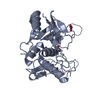
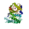
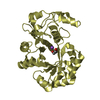
| Structure viewer | Molecule:  Molmil Molmil Jmol/JSmol Jmol/JSmol |
|---|
- Downloads & links
- Download
| PDBx/mmCIF format |  2bbm.cif.gz 2bbm.cif.gz | 73.2 KB | Display |  PDBx/mmCIF format PDBx/mmCIF format |
|---|---|---|---|---|
| PDB format |  pdb2bbm.ent.gz pdb2bbm.ent.gz | 55.2 KB | Display |  PDB format PDB format |
| PDBx/mmJSON format |  2bbm.json.gz 2bbm.json.gz | Tree view |  PDBx/mmJSON format PDBx/mmJSON format | |
| Others |  Other downloads Other downloads |
-Validation report
| Arichive directory |  https://data.pdbj.org/pub/pdb/validation_reports/bb/2bbm https://data.pdbj.org/pub/pdb/validation_reports/bb/2bbm ftp://data.pdbj.org/pub/pdb/validation_reports/bb/2bbm ftp://data.pdbj.org/pub/pdb/validation_reports/bb/2bbm | HTTPS FTP |
|---|
-Related structure data
- Links
Links
- Assembly
| Deposited unit | 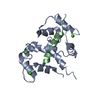
| |||||||||
|---|---|---|---|---|---|---|---|---|---|---|
| 1 |
| |||||||||
| NMR ensembles |
|
- Components
| #1: Protein | Mass: 16694.324 Da / Num. of mol.: 1 Source method: isolated from a genetically manipulated source Source: (gene. exp.)  |
|---|---|
| #2: Protein/peptide | Mass: 2972.538 Da / Num. of mol.: 1 Source method: isolated from a genetically manipulated source References: UniProt: P07313 |
| #3: Chemical | ChemComp-CA / |
-Experimental details
-Experiment
| Experiment | Method: SOLUTION NMR |
|---|
- Sample preparation
| Crystal grow | *PLUS Method: other / Details: NMR |
|---|
- Processing
| Refinement | Software ordinal: 1 Details: DETAILS OF THE STRUCTURE DETERMINATION AND ALL STRUCTURAL STATISTICS ARE GIVEN IN THE PAPER CITED ON *JRNL* RECORDS ABOVE (I.E. AGREEMENT WITH EXPERIMENTAL RESTRAINTS, DEVIATIONS FROM ...Details: DETAILS OF THE STRUCTURE DETERMINATION AND ALL STRUCTURAL STATISTICS ARE GIVEN IN THE PAPER CITED ON *JRNL* RECORDS ABOVE (I.E. AGREEMENT WITH EXPERIMENTAL RESTRAINTS, DEVIATIONS FROM IDEALITY FOR BOND LENGTHS, ANGLES, PLANES AND CHIRALITY, NON-BONDED CONTACTS, ATOMIC RMS DIFFERENCES BETWEEN THE CALCULATED STRUCTURES). THE STRUCTURES ARE BASED ON 1827 INTERPROTON DISTANCE RESTRAINTS DERIVED FROM NOE MEASUREMENTS; 148 HYDROGEN-BONDING DISTANCE RESTRAINTS FOR 74 HYDROGEN-BONDS IDENTIFIED ON THE BASIS OF THE NOE AND AMIDE PROTON EXCHANGE DATA, AS WELL AS THE INITIAL STRUCTURE CALCULATIONS; 24 RESTRAINTS FOR THE 4 CALCIUM IONS, AND 113 PHI TORSION ANGLE RESTRAINTS DERIVED FROM COUPLING DATA, CONSTANTS, NOE AND 13C SECONDARY CHEMICAL SHIFTS. THE METHOD USED TO DETERMINE THE STRUCTURES IS THE HYBRID METRIC MATRIX DISTANCE GEOMETRY-DYNAMICAL SIMULATED ANNEALING METHOD [M.NILGES, G.M.CLORE, AND A.M.GRONENBORN, FEBS LETT. 229, 317-324 (1988)]. A TOTAL OF 21 STRUCTURES WERE CALCULATED. THE COORDINATES OF THE RESTRAINED MINIMIZED STRUCTURE ARE PRESENTED IN THIS ENTRY. THIS WAS OBTAINED BY AVERAGING THE COORDINATES OF THE INDIVIDUAL STRUCTURES AND SUBJECTING THE RESULTING COORDINATES TO RESTRAINED MINIMIZATION. THE COORDINATES OF THE 21 INDIVIDUAL SA STRUCTURES ARE PRESENTED IN PROTEIN DATA BANK ENTRY 2BBN. THE LAST COLUMN IN THIS COORDINATE FILE REPRESENTS THE ATOMIC RMS DEVIATION OF THE INDIVIDUAL STRUCTURES ABOUT THE MEAN COORDINATE POSITIONS. |
|---|---|
| NMR ensemble | Conformers submitted total number: 1 |
 Movie
Movie Controller







 PDBj
PDBj


