 Movie
Movie Controller

[English] 日本語
 Yorodumi
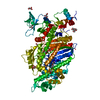
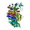
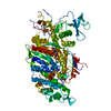
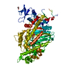
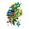
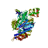
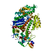
- PDB-1vom: COMPLEX BETWEEN DICTYOSTELIUM MYOSIN AND MGADP AND VANADATE AT 1.... -
+ Open data

- Basic information
| Entry | Database: PDB / ID: 1vom | ||||||
|---|---|---|---|---|---|---|---|
| Title | COMPLEX BETWEEN DICTYOSTELIUM MYOSIN AND MGADP AND VANADATE AT 1.9A RESOLUTION | ||||||
 Components Components | MYOSIN | ||||||
 Keywords Keywords | MUSCLE PROTEIN / MYOSIN / MOLECULAR MOTOR / TRANSITION-STATE ANALOG | ||||||
| Function / homology |  Function and homology information Function and homology informationuropod retraction / cytoplasmic actin-based contraction involved in forward cell motility / phagocytic cup base / pathogen-containing vacuole / response to differentiation-inducing factor 1 / equatorial cell cortex / contractile actin filament bundle assembly / pseudopodium retraction / cell trailing edge / contractile vacuole organization ...uropod retraction / cytoplasmic actin-based contraction involved in forward cell motility / phagocytic cup base / pathogen-containing vacuole / response to differentiation-inducing factor 1 / equatorial cell cortex / contractile actin filament bundle assembly / pseudopodium retraction / cell trailing edge / contractile vacuole organization / myosin filament assembly / aggregation involved in sorocarp development / culmination involved in sorocarp development / adenyl nucleotide binding / RHO GTPases activate PAKs / calcium-dependent ATPase activity / hypotonic response / actomyosin contractile ring / uropod / apical cortex / negative regulation of actin filament polymerization / actin-myosin filament sliding / detection of mechanical stimulus / substrate-dependent cell migration, cell extension / bleb assembly / actomyosin / filopodium assembly / myosin filament / early phagosome / cortical actin cytoskeleton organization / myosin II complex / cortical actin cytoskeleton / microfilament motor activity / cleavage furrow / pseudopodium / mitotic cytokinesis / cytoskeletal motor activity / response to cAMP / response to mechanical stimulus / 14-3-3 protein binding / response to hydrogen peroxide / cell motility / chemotaxis / actin filament binding / intracellular protein localization / regulation of cell shape / extracellular matrix / cytoplasmic vesicle / cell cortex / cytoskeleton / calmodulin binding / ATP binding / identical protein binding / cytoplasm / cytosol Similarity search - Function | ||||||
| Biological species |  | ||||||
| Method |  X-RAY DIFFRACTION / Resolution: 1.9 Å X-RAY DIFFRACTION / Resolution: 1.9 Å | ||||||
 Authors Authors | Rayment, I. / Smith, C.A. | ||||||
 Citation Citation | Journal: Biochemistry / Year: 1996 Title: X-ray structure of the magnesium(II).ADP.vanadate complex of the Dictyostelium discoideum myosin motor domain to 1.9 A resolution. Authors: Smith, C.A. / Rayment, I. #1: Journal: Biophys.J. / Year: 1995Title: Structural Studies of Myosin:Nucleotide Complexes: A Revised Model for the Molecular Basis of Muscle Contraction Authors: Fisher, A.J. / Smith, C.A. / Thoden, J. / Smith, R. / Sutoh, K. / Holden, H.M. / Rayment, I. #2: Journal: Biochemistry / Year: 1995Title: X-Ray Structures of the Myosin Motor Domain of Dictyostelium Discoideum Complexed with Mgadp.Befx and Mgadp.Alf4- Authors: Fisher, A.J. / Smith, C.A. / Thoden, J.B. / Smith, R. / Sutoh, K. / Holden, H.M. / Rayment, I. #3: Journal: Science / Year: 1993Title: Three-Dimensional Structure of Myosin Subfragment-1: A Molecular Motor Authors: Rayment, I. / Rypniewski, W.R. / Schmidt-Base, K. / Smith, R. / Tomchick, D.R. / Benning, M.M. / Winkelmann, D.A. / Wesenberg, G. / Holden, H.M. #4: Journal: Biochem.Biophys.Res.Commun. / Year: 1993Title: Force-Generating Domain of Myosin Motor Authors: Itakura, S. / Yamakawa, H. / Toyoshima, Y.Y. / Ishijima, A. / Kojima, T. / Harada, Y. / Yanagida, T. / Wakabayashi, T. / Sutoh, K. | ||||||
| History |
|
- Structure visualization
| Structure viewer | Molecule: MolmilJmol/JSmol |
|---|
- Downloads & links
-Download
| PDBx/mmCIF format | 1vom.cif.gz | 178.1 KB | Display | PDBx/mmCIF format |
|---|---|---|---|---|
| PDB format | pdb1vom.ent.gz | 136.2 KB | Display | PDB format |
| PDBx/mmJSON format | 1vom.json.gz | Tree view | PDBx/mmJSON format | |
| Others |  Other downloads Other downloads |
-Validation report
| Arichive directory | https://data.pdbj.org/pub/pdb/validation_reports/vo/1vomftp://data.pdbj.org/pub/pdb/validation_reports/vo/1vom | HTTPS FTP |
|---|
-Related structure data
| Similar structure data |
|---|


-Links
 PDBj
PDBj


- Assembly
| Deposited unit | 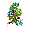
| ||||||||
|---|---|---|---|---|---|---|---|---|---|
| 1 |
| ||||||||
| Unit cell |
|
-Components
| #1: Protein | Mass: 86749.008 Da / Num. of mol.: 1 / Fragment: TRUNCATED AT RESIDUE 762 Source method: isolated from a genetically manipulated source Details: THIS MOLECULE WAS TRUNCATED TO YIELD A FRAGMENT THAT CRYSTALLIZES READILY Source: (gene. exp.) |
|---|---|
| #2: Chemical | ChemComp-MG /   Mass: 24.305 Da / Num. of mol.: 1 / Source method: obtained synthetically / Formula: Mg Mass: 24.305 Da / Num. of mol.: 1 / Source method: obtained synthetically / Formula: Mg |
| #3: Chemical | ChemComp-VO4 /   Mass: 114.939 Da / Num. of mol.: 1 / Source method: obtained synthetically / Formula: VO4 Mass: 114.939 Da / Num. of mol.: 1 / Source method: obtained synthetically / Formula: VO4 |
| #4: Chemical | ChemComp-ADP /   Mass: 427.201 Da / Num. of mol.: 1 / Source method: obtained synthetically / Formula: C10H15N5O10P2 / Comment: ADP, energy-carrying molecule*YM Mass: 427.201 Da / Num. of mol.: 1 / Source method: obtained synthetically / Formula: C10H15N5O10P2 / Comment: ADP, energy-carrying molecule*YM |
| #5: Water | ChemComp-HOH /  Mass: 18.015 Da / Num. of mol.: 705 / Source method: isolated from a natural source / Formula: H2O Mass: 18.015 Da / Num. of mol.: 705 / Source method: isolated from a natural source / Formula: H2O |
| Compound details | SSBOND THIS LIES BETWEEN TWOFOLD RELATED MOLECULES. |
-Experimental details
-Experiment
| Experiment | Method: X-RAY DIFFRACTION |
|---|
- Sample preparation
| Crystal | Density Matthews: 5.45 Å3/Da / Density % sol: 77.41 % | ||||||||||||||||||||||||||||||||||||||||||||||||||||||
|---|---|---|---|---|---|---|---|---|---|---|---|---|---|---|---|---|---|---|---|---|---|---|---|---|---|---|---|---|---|---|---|---|---|---|---|---|---|---|---|---|---|---|---|---|---|---|---|---|---|---|---|---|---|---|---|
| Crystal grow | *PLUS Temperature: 4 ℃ / pH: 7 / Method: batch method | ||||||||||||||||||||||||||||||||||||||||||||||||||||||
| Components of the solutions | *PLUS
|
-Data collection
| Diffraction source | Wavelength: 1.08 |
|---|---|
| Detector | Type: MARRESEARCH / Detector: IMAGE PLATE AREA DETECTOR / Date: Feb 1, 1995 |
| Radiation | Scattering type: x-ray |
| Radiation wavelength | Wavelength: 1.08 Å / Relative weight: 1 |
| Reflection | Num. obs: 75066 / % possible obs: 97 % / Observed criterion σ(I): 0 / Rmerge(I) obs: 0.028 |
| Reflection | *PLUS Highest resolution: 1.9 Å / Num. measured all: 393986 |
- Processing
| Software |
| ||||||||||||||||||||||||||||||
|---|---|---|---|---|---|---|---|---|---|---|---|---|---|---|---|---|---|---|---|---|---|---|---|---|---|---|---|---|---|---|---|
| Refinement | Resolution: 1.9→30 Å / σ(F): 0 /
| ||||||||||||||||||||||||||||||
| Refinement step | Cycle: LAST / Resolution: 1.9→30 Å
| ||||||||||||||||||||||||||||||
| Refine LS restraints |
| ||||||||||||||||||||||||||||||
| Software | *PLUS Name: TNT / Classification: refinement | ||||||||||||||||||||||||||||||
| Refinement | *PLUS Lowest resolution: 20 Å / Rfactor all: 0.194 | ||||||||||||||||||||||||||||||
| Solvent computation | *PLUS | ||||||||||||||||||||||||||||||
| Displacement parameters | *PLUS | ||||||||||||||||||||||||||||||
| Refine LS restraints | *PLUS
|
