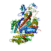
Mass: 18.015 Da / Num. of mol.: 596 / Source method: isolated from a natural source / Formula: H2O
-
Details
Nonpolymer details
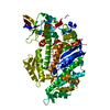
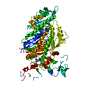
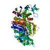
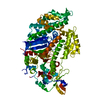
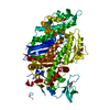
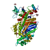
AUTHOR STATES THAT THE LONG BOND DISTANCE IN VO4 REFLECTS THAT THIS VANADATE IS A TRANSITION STATE ...AUTHOR STATES THAT THE LONG BOND DISTANCE IN VO4 REFLECTS THAT THIS VANADATE IS A TRANSITION STATE ANALOG WITH BIPYRAMIDAL GEOMETRY.IN THESE COMPLEXES THE AXIAL V-O BOND DISTANCES ARE MUCH LONGER THAN SEEN IN SIMPLE VO4 COMPLEXES.
Sequence details
THE SEQUENCE CONFLICTS ARE DUE TO VARIATIONS OF DIFFERENT VERSIONS OF THE WILD-TYPE DICTYOSTELIUM MYOSIN.
-
Experimental details
-
Experiment
Experiment
Method: X-RAY DIFFRACTION / Number of used crystals: 1
-
Sample preparation
Crystal
Density Matthews: 2.87 Å3/Da / Density % sol: 57.18 %
Crystal grow
Temperature: 278 K / Method: vapor diffusion, hanging drop / pH: 7 Details: mixing 5 ul of protein with an equal volume of well solution containing 100 mM MOPS, 250 mM MgCl2, 12% PEG 8000, 1 mM TCEP, and 2 mM Thymol, pH 7.0, vapor diffusion, hanging drop, temperature 278K
Resolution: 2.2→50 Å / Cor.coef. Fo:Fc: 0.949 / Cor.coef. Fo:Fc free: 0.915 / SU B: 5.329 / SU ML: 0.137 / Cross valid method: THROUGHOUT / ESU R: 0.222 / ESU R Free: 0.197 / Stereochemistry target values: MAXIMUM LIKELIHOOD / Details: HYDROGENS HAVE BEEN ADDED IN THE RIDING POSITIONS
Rfactor
Num. reflection
% reflection
Selection details
Rfree
0.24479
2569
5.1 %
RANDOM
Rwork
0.19224
-
-
-
obs
0.19493
48034
99.11 %
-
Solvent computation
Ion probe radii: 0.8 Å / Shrinkage radii: 0.8 Å / VDW probe radii: 1.2 Å / Solvent model: MASK
Displacement parameters
Biso mean: 34.536 Å2
Baniso -1
Baniso -2
Baniso -3
1-
-0.3 Å2
0 Å2
0 Å2
2-
-
2.7 Å2
0 Å2
3-
-
-
-2.4 Å2
Refinement step
Cycle: LAST / Resolution: 2.2→50 Å
Protein
Nucleic acid
Ligand
Solvent
Total
Num. atoms
5528
0
60
596
6184
Refine LS restraints
Refine-ID
Type
Dev ideal
Dev ideal target
Number
X-RAY DIFFRACTION
r_bond_refined_d
0.022
0.022
5702
X-RAY DIFFRACTION
r_bond_other_d
X-RAY DIFFRACTION
r_angle_refined_deg
1.826
1.959
7722
X-RAY DIFFRACTION
r_angle_other_deg
X-RAY DIFFRACTION
r_dihedral_angle_1_deg
6.278
5
708
X-RAY DIFFRACTION
r_dihedral_angle_2_deg
36.685
24.8
275
X-RAY DIFFRACTION
r_dihedral_angle_3_deg
15.307
15
951
X-RAY DIFFRACTION
r_dihedral_angle_4_deg
18.489
15
28
X-RAY DIFFRACTION
r_chiral_restr
0.123
0.2
848
X-RAY DIFFRACTION
r_gen_planes_refined
0.008
0.02
4349
X-RAY DIFFRACTION
r_gen_planes_other
X-RAY DIFFRACTION
r_nbd_refined
0.211
0.2
2651
X-RAY DIFFRACTION
r_nbd_other
X-RAY DIFFRACTION
r_nbtor_refined
0.307
0.2
3899
X-RAY DIFFRACTION
r_nbtor_other
X-RAY DIFFRACTION
r_xyhbond_nbd_refined
0.207
0.2
503
X-RAY DIFFRACTION
r_xyhbond_nbd_other
X-RAY DIFFRACTION
r_metal_ion_refined
0.323
0.2
2
X-RAY DIFFRACTION
r_metal_ion_other
X-RAY DIFFRACTION
r_symmetry_vdw_refined
0.226
0.2
46
X-RAY DIFFRACTION
r_symmetry_vdw_other
X-RAY DIFFRACTION
r_symmetry_hbond_refined
0.299
0.2
13
X-RAY DIFFRACTION
r_symmetry_hbond_other
X-RAY DIFFRACTION
r_symmetry_metal_ion_refined
X-RAY DIFFRACTION
r_symmetry_metal_ion_other
X-RAY DIFFRACTION
r_mcbond_it
1.147
1.5
3647
X-RAY DIFFRACTION
r_mcbond_other
X-RAY DIFFRACTION
r_mcangle_it
1.855
2
5639
X-RAY DIFFRACTION
r_scbond_it
2.817
3
2389
X-RAY DIFFRACTION
r_scangle_it
4
4.5
2081
X-RAY DIFFRACTION
r_rigid_bond_restr
X-RAY DIFFRACTION
r_sphericity_free
X-RAY DIFFRACTION
r_sphericity_bonded
LS refinement shell
Resolution: 2.2→2.256 Å / Total num. of bins used: 20
Rfactor
Num. reflection
% reflection
Rfree
0.326
167
-
Rwork
0.234
3332
-
obs
-
-
93.91 %
+
About Yorodumi
-
News
-
Feb 9, 2022. New format data for meta-information of EMDB entries
New format data for meta-information of EMDB entries
Version 3 of the EMDB header file is now the official format.
The previous official version 1.9 will be removed from the archive.
In the structure databanks used in Yorodumi, some data are registered as the other names, "COVID-19 virus" and "2019-nCoV". Here are the details of the virus and the list of structure data.
Jan 31, 2019. EMDB accession codes are about to change! (news from PDBe EMDB page)
EMDB accession codes are about to change! (news from PDBe EMDB page)
The allocation of 4 digits for EMDB accession codes will soon come to an end. Whilst these codes will remain in use, new EMDB accession codes will include an additional digit and will expand incrementally as the available range of codes is exhausted. The current 4-digit format prefixed with “EMD-” (i.e. EMD-XXXX) will advance to a 5-digit format (i.e. EMD-XXXXX), and so on. It is currently estimated that the 4-digit codes will be depleted around Spring 2019, at which point the 5-digit format will come into force.
The EM Navigator/Yorodumi systems omit the EMD- prefix.
Related info.:Q: What is EMD? / ID/Accession-code notation in Yorodumi/EM Navigator
Yorodumi is a browser for structure data from EMDB, PDB, SASBDB, etc.
This page is also the successor to EM Navigator detail page, and also detail information page/front-end page for Omokage search.
The word "yorodu" (or yorozu) is an old Japanese word meaning "ten thousand". "mi" (miru) is to see.
Related info.:EMDB / PDB / SASBDB / Comparison of 3 databanks / Yorodumi Search / Aug 31, 2016. New EM Navigator & Yorodumi / Yorodumi Papers / Jmol/JSmol / Function and homology information / Changes in new EM Navigator and Yorodumi
 Movie
Movie Controller

 Yorodumi
 Open data
 Basic information
 Components
 Keywords
 Function and homology information
 X-RAY DIFFRACTION /
  Authors
 Citation
 Structure visualization
 Downloads & links
 Other downloads
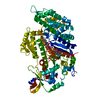


 PDBj
PDBj


 Assembly


 Mass: 24.305 Da / Num. of mol.: 2 / Source method: obtained synthetically / Formula: Mg
 Mass: 114.939 Da / Num. of mol.: 1 / Source method: obtained synthetically / Formula: VO4
 Mass: 427.201 Da / Num. of mol.: 1 / Source method: obtained synthetically / Formula: C10H15N5O10P2 / Comment: ADP, energy-carrying molecule*YM
 Mass: 62.068 Da / Num. of mol.: 1 / Source method: obtained synthetically / Formula: C2H6O2
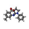
 Mass: 292.332 Da / Num. of mol.: 1 / Source method: obtained synthetically / Formula: C18H16N2O2
 Sample preparation
 / Beamline: 32-ID / Wavelength: 1 Å
 Processing