 Movie
Movie Controller

[English] 日本語
 Yorodumi
- PDB-6due: Toxoplasma gondii MyoA, a Class-XIV myosin, in the pre-powerstrok... -
+ Open data

- Basic information
| Entry | Database: PDB / ID: 6due | ||||||
|---|---|---|---|---|---|---|---|
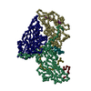
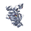
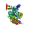
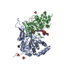
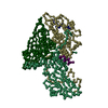
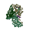
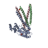
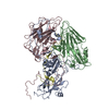
| Title | Toxoplasma gondii MyoA, a Class-XIV myosin, in the pre-powerstroke state | ||||||
 Components Components | Myosin A | ||||||
 Keywords Keywords | MOTOR PROTEIN / Apicomplexan / Myosin / ATPase | ||||||
| Function / homology |  Function and homology information Function and homology informationmyosin complex / cytoskeletal motor activity / actin binding / ATP binding / metal ion binding Similarity search - Function | ||||||
| Biological species |  | ||||||
| Method |  X-RAY DIFFRACTION / SYNCHROTRON / MOLECULAR REPLACEMENT / molecular replacement / Resolution: 2.6 Å X-RAY DIFFRACTION / SYNCHROTRON / MOLECULAR REPLACEMENT / molecular replacement / Resolution: 2.6 Å | ||||||
 Authors Authors | Powell, C.J. / Boulanger, M.J. | ||||||
| Funding support |  Canada, 1items Canada, 1items
| ||||||
 Citation Citation | Journal: Proc. Natl. Acad. Sci. U.S.A. / Year: 2018 Title: Structural and mechanistic insights into the function of the unconventional class XIV myosin MyoA fromToxoplasma gondii. Authors: Powell, C.J. / Ramaswamy, R. / Kelsen, A. / Hamelin, D.J. / Warshaw, D.M. / Bosch, J. / Burke, J.E. / Ward, G.E. / Boulanger, M.J. | ||||||
| History |
|
- Structure visualization
| Structure viewer | Molecule: MolmilJmol/JSmol |
|---|
- Downloads & links
-Download
| PDBx/mmCIF format | 6due.cif.gz | 311.3 KB | Display | PDBx/mmCIF format |
|---|---|---|---|---|
| PDB format | pdb6due.ent.gz | 249 KB | Display | PDB format |
| PDBx/mmJSON format | 6due.json.gz | Tree view | PDBx/mmJSON format | |
| Others |  Other downloads Other downloads |
-Validation report
| Arichive directory | https://data.pdbj.org/pub/pdb/validation_reports/du/6dueftp://data.pdbj.org/pub/pdb/validation_reports/du/6due | HTTPS FTP |
|---|
-Related structure data
| Related structure data |  4byfS S: Starting model for refinement |
|---|---|
| Similar structure data |

-Links
 PDBj
PDBj


- Assembly
| Deposited unit | 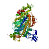
| ||||||||
|---|---|---|---|---|---|---|---|---|---|
| 1 |
| ||||||||
| Unit cell |
|
-Components
-Protein , 1 types, 1 molecules A
| #1: Protein | Mass: 88298.398 Da / Num. of mol.: 1 Source method: isolated from a genetically manipulated source Source: (gene. exp.) Strain: ATCC 50853 / GT1 / Gene: TGGT1_235470 / Production host:   Spodoptera frugiperda (fall armyworm) / References: UniProt: S7W634 Spodoptera frugiperda (fall armyworm) / References: UniProt: S7W634 |
|---|
-Non-polymers , 5 types, 31 molecules 


| #2: Chemical | ChemComp-ADP /  Mass: 427.201 Da / Num. of mol.: 1 / Source method: obtained synthetically / Formula: C10H15N5O10P2 / Comment: ADP, energy-carrying molecule*YM Mass: 427.201 Da / Num. of mol.: 1 / Source method: obtained synthetically / Formula: C10H15N5O10P2 / Comment: ADP, energy-carrying molecule*YM | ||
|---|---|---|---|
| #3: Chemical | ChemComp-ALF /  Mass: 102.975 Da / Num. of mol.: 1 / Source method: obtained synthetically / Formula: AlF4 Mass: 102.975 Da / Num. of mol.: 1 / Source method: obtained synthetically / Formula: AlF4 | ||
| #4: Chemical | ChemComp-MG /  Mass: 24.305 Da / Num. of mol.: 1 / Source method: obtained synthetically / Formula: Mg Mass: 24.305 Da / Num. of mol.: 1 / Source method: obtained synthetically / Formula: Mg | ||
| #5: Chemical |  Mass: 92.094 Da / Num. of mol.: 3 / Source method: obtained synthetically / Formula: C3H8O3 Mass: 92.094 Da / Num. of mol.: 3 / Source method: obtained synthetically / Formula: C3H8O3#6: Water | ChemComp-HOH / | Mass: 18.015 Da / Num. of mol.: 25 / Source method: isolated from a natural source / Formula: H2O |
-Experimental details
-Experiment
| Experiment | Method: X-RAY DIFFRACTION / Number of used crystals: 1 |
|---|
- Sample preparation
| Crystal | Density Matthews: 2.32 Å3/Da / Density % sol: 47.08 % |
|---|---|
| Crystal grow | Temperature: 277 K / Method: vapor diffusion, sitting drop Details: 1:1 protein solution to reservoir solution (22% PEG8000, 200 mM ammonium sulfate, 100 mM MES, pH 6.5), cryoprotectant: reservoir solution + 15% glycerol PH range: 6.5-7.5 |
-Data collection
| Diffraction | Mean temperature: 100 K |
|---|---|
| Diffraction source | Source: SYNCHROTRON / Site: SSRL  / Beamline: BL14-1 / Wavelength: 0.987 Å / Beamline: BL14-1 / Wavelength: 0.987 Å |
| Detector | Type: MARMOSAIC 325 mm CCD / Detector: CCD / Date: Jul 20, 2017 Details: Flat bent collimating Rh coated mirror, toroidal focussing mirror |
| Radiation | Monochromator: double crystal Si(111) / Protocol: SINGLE WAVELENGTH / Monochromatic (M) / Laue (L): M / Scattering type: x-ray |
| Radiation wavelength | Wavelength: 0.987 Å / Relative weight: 1 |
| Reflection | Resolution: 2.6→85.95 Å / Num. obs: 24306 / % possible obs: 97.6 % / Redundancy: 3.8 % / Biso Wilson estimate: 49.28 Å2 / Rmerge(I) obs: 0.124 / Net I/σ(I): 7.3 |
| Reflection shell | Resolution: 2.6→2.74 Å / Redundancy: 3.9 % / Rmerge(I) obs: 0.856 / Mean I/σ(I) obs: 1.6 / Num. unique obs: 3495 / % possible all: 96.7 |
-Phasing
| Phasing | Method: molecular replacement |
|---|
- Processing
| Software |
| ||||||||||||||||||||||||||||||||||||||||||||||||||||||||||||||||||||||
|---|---|---|---|---|---|---|---|---|---|---|---|---|---|---|---|---|---|---|---|---|---|---|---|---|---|---|---|---|---|---|---|---|---|---|---|---|---|---|---|---|---|---|---|---|---|---|---|---|---|---|---|---|---|---|---|---|---|---|---|---|---|---|---|---|---|---|---|---|---|---|---|
| Refinement | Method to determine structure: MOLECULAR REPLACEMENT Starting model: PDB entry 4BYF Resolution: 2.6→48.717 Å / SU ML: 0.39 / Cross valid method: THROUGHOUT / σ(F): 1.09 / Phase error: 31.13
| ||||||||||||||||||||||||||||||||||||||||||||||||||||||||||||||||||||||
| Solvent computation | Shrinkage radii: 0.9 Å / VDW probe radii: 1.11 Å | ||||||||||||||||||||||||||||||||||||||||||||||||||||||||||||||||||||||
| Displacement parameters | Biso max: 180.27 Å2 / Biso mean: 68.7476 Å2 / Biso min: 28.04 Å2 | ||||||||||||||||||||||||||||||||||||||||||||||||||||||||||||||||||||||
| Refinement step | Cycle: final / Resolution: 2.6→48.717 Å
| ||||||||||||||||||||||||||||||||||||||||||||||||||||||||||||||||||||||
| LS refinement shell | Refine-ID: X-RAY DIFFRACTION / Rfactor Rfree error: 0 / Total num. of bins used: 9
| ||||||||||||||||||||||||||||||||||||||||||||||||||||||||||||||||||||||
| Refinement TLS params. | Method: refined / Origin x: 20.9759 Å / Origin y: 2.0842 Å / Origin z: 21.2636 Å
| ||||||||||||||||||||||||||||||||||||||||||||||||||||||||||||||||||||||
| Refinement TLS group |
|
