 Movie
Movie Controller


+ Open data

- Basic information
| Entry | Database: PDB / ID: 6aj0 | ||||||||||||||||||||||||||||||||||||||||||||||||||||||
|---|---|---|---|---|---|---|---|---|---|---|---|---|---|---|---|---|---|---|---|---|---|---|---|---|---|---|---|---|---|---|---|---|---|---|---|---|---|---|---|---|---|---|---|---|---|---|---|---|---|---|---|---|---|---|---|
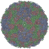
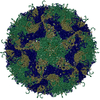
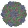
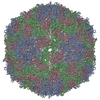
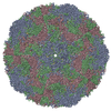
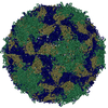
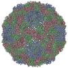
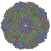
| Title | The structure of Enterovirus D68 mature virion | ||||||||||||||||||||||||||||||||||||||||||||||||||||||
 Components Components |
| ||||||||||||||||||||||||||||||||||||||||||||||||||||||
 Keywords Keywords | VIRUS / Enterovirus D68 / mature virion | ||||||||||||||||||||||||||||||||||||||||||||||||||||||
| Function / homology |  Function and homology information Function and homology informationhost cell membrane / cysteine-type peptidase activity / helicase activity / picornain 2A / symbiont-mediated suppression of host mRNA export from nucleus / symbiont genome entry into host cell via pore formation in plasma membrane / picornain 3C / T=pseudo3 icosahedral viral capsid / host cell cytoplasmic vesicle membrane / viral capsid ...host cell membrane / cysteine-type peptidase activity / helicase activity / picornain 2A / symbiont-mediated suppression of host mRNA export from nucleus / symbiont genome entry into host cell via pore formation in plasma membrane / picornain 3C / T=pseudo3 icosahedral viral capsid / host cell cytoplasmic vesicle membrane / viral capsid / ribonucleoside triphosphate phosphatase activity / host cell / nucleoside-triphosphate phosphatase / channel activity / monoatomic ion transmembrane transport / host cell cytoplasm / RNA helicase activity / symbiont-mediated suppression of host innate immune response / endocytosis involved in viral entry into host cell / symbiont-mediated suppression of host gene expression / symbiont-mediated activation of host autophagy / RNA-directed RNA polymerase / cysteine-type endopeptidase activity / viral RNA genome replication / RNA-directed RNA polymerase activity / symbiont entry into host cell / virion attachment to host cell / host cell nucleus / structural molecule activity / DNA-templated transcription / proteolysis / RNA binding / zinc ion binding / ATP binding Similarity search - Function | ||||||||||||||||||||||||||||||||||||||||||||||||||||||
| Biological species |  Enterovirus D68 Enterovirus D68 | ||||||||||||||||||||||||||||||||||||||||||||||||||||||
| Method | ELECTRON MICROSCOPY / single particle reconstruction / cryo EM / Resolution: 3.4 Å | ||||||||||||||||||||||||||||||||||||||||||||||||||||||
 Authors Authors | Zheng, Q.B. / Zhu, R. / Xu, L.F. / He, M.Z. / Yan, X.D. / Cheng, T. / Li, S.W. | ||||||||||||||||||||||||||||||||||||||||||||||||||||||
| Funding support |  China, China,  United States, 11items United States, 11items
| ||||||||||||||||||||||||||||||||||||||||||||||||||||||
 Citation Citation | Journal: Nat Microbiol / Year: 2019 Title: Atomic structures of enterovirus D68 in complex with two monoclonal antibodies define distinct mechanisms of viral neutralization. Authors: Qingbing Zheng / Rui Zhu / Longfa Xu / Maozhou He / Xiaodong Yan / Dongxiao Liu / Zhichao Yin / Yangtao Wu / Yongchao Li / Lisheng Yang / Wangheng Hou / Shuxuan Li / Zizhen Li / Zhenqin Chen ...Authors: Qingbing Zheng / Rui Zhu / Longfa Xu / Maozhou He / Xiaodong Yan / Dongxiao Liu / Zhichao Yin / Yangtao Wu / Yongchao Li / Lisheng Yang / Wangheng Hou / Shuxuan Li / Zizhen Li / Zhenqin Chen / Zhihai Li / Hai Yu / Ying Gu / Jun Zhang / Timothy S Baker / Z Hong Zhou / Barney S Graham / Tong Cheng / Shaowei Li / Ningshao Xia / Abstract: Enterovirus D68 (EV-D68) undergoes structural transformation between mature, cell-entry intermediate (A-particle) and empty forms throughout its life cycle. Structural information for the various ...Enterovirus D68 (EV-D68) undergoes structural transformation between mature, cell-entry intermediate (A-particle) and empty forms throughout its life cycle. Structural information for the various forms and antibody-bound capsids will facilitate the development of effective vaccines and therapeutics against EV-D68 infection, which causes childhood respiratory and paralytic diseases worldwide. Here, we report the structures of three EV-D68 capsid states representing the virus at major phases. We further describe two original monoclonal antibodies (15C5 and 11G1) with distinct structurally defined mechanisms for virus neutralization. 15C5 and 11G1 engage the capsid loci at icosahedral three-fold and five-fold axes, respectively. To block viral attachment, 15C5 binds three forms of capsids, and triggers mature virions to transform into A-particles, mimicking engagement by the functional receptor ICAM-5, whereas 11G1 exclusively recognizes the A-particle. Our data provide a structural and molecular explanation for the transition of picornavirus capsid conformations and demonstrate distinct mechanisms for antibody-mediated neutralization. | ||||||||||||||||||||||||||||||||||||||||||||||||||||||
| History |
|
- Structure visualization
| Movie |
Movie viewer |
|---|---|
| Structure viewer | Molecule: MolmilJmol/JSmol |
- Downloads & links
-Download
| PDBx/mmCIF format | 6aj0.cif.gz | 151.9 KB | Display | PDBx/mmCIF format |
|---|---|---|---|---|
| PDB format | pdb6aj0.ent.gz | 117.1 KB | Display | PDB format |
| PDBx/mmJSON format | 6aj0.json.gz | Tree view | PDBx/mmJSON format | |
| Others |  Other downloads Other downloads |
-Validation report
| Arichive directory | https://data.pdbj.org/pub/pdb/validation_reports/aj/6aj0ftp://data.pdbj.org/pub/pdb/validation_reports/aj/6aj0 | HTTPS FTP |
|---|
-Related structure data
| Related structure data | 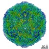 9629MC 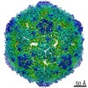 9631C 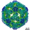 9632C 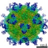 9633C 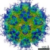 9634C 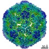 9635C 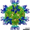 9636C 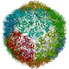 6aj2C  6aj3C 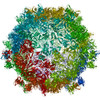 6aj7C 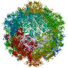 6aj9C M: map data used to model this data C: citing same article ( |
|---|---|
| Similar structure data |
-Links
 PDBj
PDBj

- Assembly
| Deposited unit | 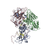
|
|---|---|
| 1 | x 60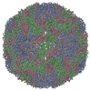
|
| 2 |
|
| 3 | x 5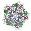
|
| 4 | x 6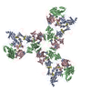
|
| 5 | 
|
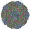
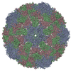
| Symmetry | Point symmetry: (Schoenflies symbol: I (icosahedral)) |
-Components
| #1: Protein | Mass: 32673.035 Da / Num. of mol.: 1 / Source method: isolated from a natural source / Source: (natural) Enterovirus D68 / References: UniProt: A0A097F8Q2 |
|---|---|
| #2: Protein | Mass: 27567.135 Da / Num. of mol.: 1 / Source method: isolated from a natural source / Source: (natural) Enterovirus D68 / References: UniProt: A0A0A7X639, UniProt: A0A097F8Q2*PLUS |
| #3: Protein | Mass: 27142.842 Da / Num. of mol.: 1 / Source method: isolated from a natural source / Source: (natural) Enterovirus D68 / References: UniProt: A0A097F8Q2*PLUS |
| #4: Protein | Mass: 7468.157 Da / Num. of mol.: 1 / Source method: isolated from a natural source / Source: (natural) Enterovirus D68 / References: UniProt: E7FM39, UniProt: A0A097F8Q2*PLUS |
| Has protein modification | N |
-Experimental details
-Experiment
| Experiment | Method: ELECTRON MICROSCOPY |
|---|---|
| EM experiment | Aggregation state: PARTICLE / 3D reconstruction method: single particle reconstruction |
- Sample preparation
| Component | Name: Coxsackievirus A10 / Type: VIRUS / Entity ID: all / Source: NATURAL |
|---|---|
| Source (natural) | Organism: 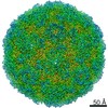 Coxsackievirus A10 Coxsackievirus A10 |
| Details of virus | Empty: NO / Enveloped: NO / Isolate: STRAIN / Type: VIRION |
| Buffer solution | pH: 7.4 |
| Specimen | Embedding applied: NO / Shadowing applied: NO / Staining applied: NO / Vitrification applied: YES |
| Vitrification | Cryogen name: ETHANE |
- Electron microscopy imaging
| Experimental equipment |  Model: Tecnai F30 / Image courtesy: FEI Company |
|---|---|
| Microscopy | Model: FEI TECNAI F30 |
| Electron gun | Electron source:  FIELD EMISSION GUN / Accelerating voltage: 300 kV / Illumination mode: FLOOD BEAM FIELD EMISSION GUN / Accelerating voltage: 300 kV / Illumination mode: FLOOD BEAM |
| Electron lens | Mode: BRIGHT FIELD |
| Image recording | Electron dose: 25 e/Å2 / Film or detector model: FEI FALCON II (4k x 4k) |
- Processing
| Software | Name: PHENIX / Version: 1.11.1_2575: / Classification: refinement | ||||||||||||||||||||||||
|---|---|---|---|---|---|---|---|---|---|---|---|---|---|---|---|---|---|---|---|---|---|---|---|---|---|
| EM software |
| ||||||||||||||||||||||||
| CTF correction | Type: PHASE FLIPPING ONLY | ||||||||||||||||||||||||
| Symmetry | Point symmetry: I (icosahedral) | ||||||||||||||||||||||||
| 3D reconstruction | Resolution: 3.4 Å / Resolution method: FSC 0.143 CUT-OFF / Num. of particles: 11938 / Symmetry type: POINT | ||||||||||||||||||||||||
| Refine LS restraints |
|
