 Movie
Movie Controller

[English] 日本語
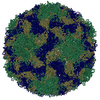
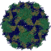
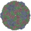
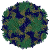
 Yorodumi
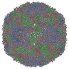
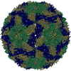
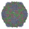
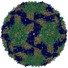
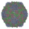
- PDB-5apm: Multiple capsid-stabilizing protein-RNA and protein-protein inter... -
+ Open data

- Basic information
| Entry | Database: PDB / ID: 5apm | ||||||
|---|---|---|---|---|---|---|---|
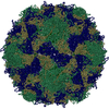
| Title | Multiple capsid-stabilizing protein-RNA and protein-protein interactions revealed in a high-resolution structure of an emerging picornavirus causing neonatal sepsis | ||||||
 Components Components |
| ||||||
 Keywords Keywords | VIRUS / PICORNAVIRUS / PARECHOVIRUS / HUMAN PARECHOVIRUS 3 / HPEV3 / NEONATAL SEPSIS / CRYOEM / IMAGE PROCESSING / SINGLE PARTICLE ANALYSIS | ||||||
| Function / homology |  Function and homology information Function and homology informationT=pseudo3 icosahedral viral capsid / host cell cytoplasmic vesicle membrane / channel activity / monoatomic ion transmembrane transport / RNA helicase activity / cysteine-type endopeptidase activity / viral RNA genome replication / RNA-directed RNA polymerase activity / symbiont entry into host cell / DNA-templated transcription ...T=pseudo3 icosahedral viral capsid / host cell cytoplasmic vesicle membrane / channel activity / monoatomic ion transmembrane transport / RNA helicase activity / cysteine-type endopeptidase activity / viral RNA genome replication / RNA-directed RNA polymerase activity / symbiont entry into host cell / DNA-templated transcription / virion attachment to host cell / structural molecule activity / proteolysis / RNA binding / ATP binding Similarity search - Function | ||||||
| Biological species |  HUMAN PARECHOVIRUS 3 HUMAN PARECHOVIRUS 3 | ||||||
| Method | ELECTRON MICROSCOPY / single particle reconstruction / Resolution: 4.3 Å | ||||||
 Authors Authors | Shakeel, S. / Westerhuis, B.M. / Domanska, A. / Koning, R.I. / Matadeen, R. / Koster, A.J. / Bakker, A.Q. / Beaumont, T. / Wolthers, K.C. / Butcher, S.J. | ||||||
 Citation Citation | Journal: Nat Commun / Year: 2016 Title: Multiple capsid-stabilizing interactions revealed in a high-resolution structure of an emerging picornavirus causing neonatal sepsis. Authors: Shabih Shakeel / Brenda M Westerhuis / Ausra Domanska / Roman I Koning / Rishi Matadeen / Abraham J Koster / Arjen Q Bakker / Tim Beaumont / Katja C Wolthers / Sarah J Butcher /   Abstract: The poorly studied picornavirus, human parechovirus 3 (HPeV3) causes neonatal sepsis with no therapies available. Our 4.3-Å resolution structure of HPeV3 on its own and at 15 Å resolution in ...The poorly studied picornavirus, human parechovirus 3 (HPeV3) causes neonatal sepsis with no therapies available. Our 4.3-Å resolution structure of HPeV3 on its own and at 15 Å resolution in complex with human monoclonal antibody Fabs demonstrates the expected picornavirus capsid structure with three distinct features. First, 25% of the HPeV3 RNA genome in 60 sites is highly ordered as confirmed by asymmetric reconstruction, and interacts with conserved regions of the capsid proteins VP1 and VP3. Second, the VP0 N terminus stabilizes the capsid inner surface, in contrast to other picornaviruses where on expulsion as VP4, it forms an RNA translocation channel. Last, VP1's hydrophobic pocket, the binding site for the antipicornaviral drug, pleconaril, is blocked and thus inappropriate for antiviral development. Together, these results suggest a direction for development of neutralizing antibodies, antiviral drugs based on targeting the RNA-protein interactions and dissection of virus assembly on the basis of RNA nucleation. | ||||||
| History |
|
- Structure visualization
| Movie |
Movie viewer |
|---|---|
| Structure viewer | Molecule: MolmilJmol/JSmol |
- Downloads & links
-Download
| PDBx/mmCIF format | 5apm.cif.gz | 139.5 KB | Display | PDBx/mmCIF format |
|---|---|---|---|---|
| PDB format | pdb5apm.ent.gz | 100.8 KB | Display | PDB format |
| PDBx/mmJSON format | 5apm.json.gz | Tree view | PDBx/mmJSON format | |
| Others |  Other downloads Other downloads |
-Validation report
| Arichive directory | https://data.pdbj.org/pub/pdb/validation_reports/ap/5apmftp://data.pdbj.org/pub/pdb/validation_reports/ap/5apm | HTTPS FTP |
|---|
-Related structure data
| Related structure data | 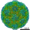 3137MC 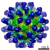 3138C 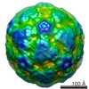 3322C M: map data used to model this data C: citing same article ( |
|---|---|
| Similar structure data | |
| EM raw data | EMPIAR-10033 (Title: Multiple capsid-stabilizing protein-RNA and protein-protein interactions revealed in a high-resolution structure of an emerging picornavirus causing neonatal sepsis Data size: 3.3 TB Data #1: Selected aligned movie frames, aligned-averaged micrographs and particle coordinates of human parechovirus 3 (HPeV3) virions [Aligned movie frames (.mrcs extension), aligned-averaged ...Data #1: Selected aligned movie frames, aligned-averaged micrographs and particle coordinates of human parechovirus 3 (HPeV3) virions [Aligned movie frames (.mrcs extension), aligned-averaged micrographs (.mrc extension) and particle coordinates (.box extension)] Data #2: Unaligned movie frames of human parechovirus 3 (HPeV3) virions [micrographs - multiframe]) |
-Links
 PDBj
PDBj

- Assembly
| Deposited unit | 
|
|---|---|
| 1 | x 60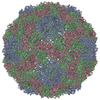
|
| 2 |
|
| 3 | x 5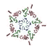
|
| 4 | x 6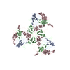
|
| 5 | 
|
| Symmetry | Point symmetry: (Schoenflies symbol: I (icosahedral)) |
-Components
| #1: Protein | Mass: 22393.314 Da / Num. of mol.: 1 / Source method: isolated from a natural source / Source: (natural) HUMAN PARECHOVIRUS 3 / Cell line: VERO / Variant: 152037 / References: UniProt: D2IE17 |
|---|---|
| #2: Protein | Mass: 26663.111 Da / Num. of mol.: 1 / Source method: isolated from a natural source / Source: (natural) HUMAN PARECHOVIRUS 3 / Cell line: VERO / Variant: 152037 / References: UniProt: D2IE17 |
| #3: Protein | Mass: 28943.061 Da / Num. of mol.: 1 / Source method: isolated from a natural source / Source: (natural) HUMAN PARECHOVIRUS 3 / Cell line: VERO / Variant: 152037 / References: UniProt: D2IE17 |
| Sequence details | WE MODELLED RESIDUES 24-221. WE MODELLED RESIDUES 20-256. WE MODELLED RESIDUES 20-282. |
-Experimental details
-Experiment
| Experiment | Method: ELECTRON MICROSCOPY |
|---|---|
| EM experiment | Aggregation state: PARTICLE / 3D reconstruction method: single particle reconstruction |
- Sample preparation
| Component | Name: HUMAN PARECHOVIRUS 3 / Type: VIRUS / Details: FORMALDEHYDE-INACTIVATED VIRUS WAS IMAGED. |
|---|---|
| Buffer solution | Name: 10MM TRIS-HCL, 150MM NACL, 1MM MGCL2 / pH: 7.5 / Details: 10MM TRIS-HCL, 150MM NACL, 1MM MGCL2 |
| Specimen | Conc.: 1 mg/ml / Embedding applied: NO / Shadowing applied: NO / Staining applied: NO / Vitrification applied: NO |
| Specimen support | Details: HOLEY CARBON |
| Vitrification | Instrument: LEICA EM GP / Cryogen name: ETHANE / Details: LIQUID ETHANE |
- Electron microscopy imaging
| Experimental equipment |  Model: Titan Krios / Image courtesy: FEI Company |
|---|---|
| Microscopy | Model: FEI TITAN KRIOS / Date: Jan 21, 2015 |
| Electron gun | Electron source:  FIELD EMISSION GUN / Accelerating voltage: 300 kV / Illumination mode: FLOOD BEAM FIELD EMISSION GUN / Accelerating voltage: 300 kV / Illumination mode: FLOOD BEAM |
| Electron lens | Mode: BRIGHT FIELD / Nominal magnification: 59000 X / Nominal defocus max: 2340 nm / Nominal defocus min: 420 nm / Cs: 0.01 mm |
| Image recording | Electron dose: 36 e/Å2 / Film or detector model: FEI FALCON II (4k x 4k) |
| Image scans | Num. digital images: 1028 |
- Processing
| EM software |
| ||||||||||||||||||||||||||||||||
|---|---|---|---|---|---|---|---|---|---|---|---|---|---|---|---|---|---|---|---|---|---|---|---|---|---|---|---|---|---|---|---|---|---|
| CTF correction | Details: MICROGRAPHS | ||||||||||||||||||||||||||||||||
| Symmetry | Point symmetry: I (icosahedral) | ||||||||||||||||||||||||||||||||
| 3D reconstruction | Method: MAXIMUM LIKELIHOOD METHOD. / Resolution: 4.3 Å / Num. of particles: 8889 / Nominal pixel size: 1.14 Å Details: WE DID NOT MODELLED DISORDERED REGIONS. THE PROVIDE COORDINATES ARE FOR AN ASYMMETRIC UNIT OF THE VIRUS. IN ORDER TO GENERATE THE WHOLE CAPSID, PLEASE USE THE MATRICES PROVIDED IN REMARK 350. ...Details: WE DID NOT MODELLED DISORDERED REGIONS. THE PROVIDE COORDINATES ARE FOR AN ASYMMETRIC UNIT OF THE VIRUS. IN ORDER TO GENERATE THE WHOLE CAPSID, PLEASE USE THE MATRICES PROVIDED IN REMARK 350. SUBMISSION BASED ON EXPERIMENTAL DATA FROM EMDB EMD-3137. (DEPOSITION ID: 13643). Symmetry type: POINT | ||||||||||||||||||||||||||||||||
| Atomic model building | Protocol: OTHER / Space: REAL / Target criteria: Cross-correlation coefficient Details: METHOD--LOCAL CORRELATION REFINEMENT PROTOCOL--HOMOLOGY MODEL | ||||||||||||||||||||||||||||||||
| Refinement | Highest resolution: 4.3 Å | ||||||||||||||||||||||||||||||||
| Refinement step | Cycle: LAST / Highest resolution: 4.3 Å
|
