 Movie
Movie Controller

[English] 日本語
 Yorodumi
- PDB-4byf: Crystal structure of human Myosin 1c in complex with calmodulin i... -
+ Open data

- Basic information
| Entry | Database: PDB / ID: 4byf | ||||||
|---|---|---|---|---|---|---|---|
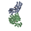
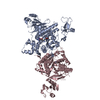
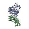
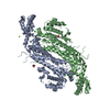
| Title | Crystal structure of human Myosin 1c in complex with calmodulin in the pre-power stroke state | ||||||
 Components Components |
| ||||||
 Keywords Keywords | HYDROLASE / MYO1C / GLUT4 EXOCYTOSIS / ATPASE / MOTOR PROTEIN | ||||||
| Function / homology |  Function and homology information Function and homology informationstereocilium membrane / positive regulation of cellular response to insulin stimulus / vesicle transport along actin filament / unconventional myosin complex / : / : / : / : / positive regulation of protein autophosphorylation / : ...stereocilium membrane / positive regulation of cellular response to insulin stimulus / vesicle transport along actin filament / unconventional myosin complex / : / : / : / : / positive regulation of protein autophosphorylation / : / negative regulation of peptidyl-threonine phosphorylation / B-WICH complex / actin filament-based movement / : / type 3 metabotropic glutamate receptor binding / Sensory processing of sound by outer hair cells of the cochlea / positive regulation of DNA binding / protein targeting to membrane / CaM pathway / Cam-PDE 1 activation / Sensory processing of sound by inner hair cells of the cochlea / positive regulation of peptidyl-threonine phosphorylation / Sodium/Calcium exchangers / positive regulation of transcription by RNA polymerase III / regulation of bicellular tight junction assembly / Calmodulin induced events / Reduction of cytosolic Ca++ levels / vascular endothelial growth factor signaling pathway / Activation of Ca-permeable Kainate Receptor / CREB1 phosphorylation through the activation of CaMKII/CaMKK/CaMKIV cascasde / Loss of phosphorylation of MECP2 at T308 / CREB1 phosphorylation through the activation of Adenylate Cyclase / negative regulation of high voltage-gated calcium channel activity / PKA activation / CaMK IV-mediated phosphorylation of CREB / Glycogen breakdown (glycogenolysis) / microfilament motor activity / response to corticosterone / negative regulation of ryanodine-sensitive calcium-release channel activity / Activation of RAC1 downstream of NMDARs / organelle localization by membrane tethering / CLEC7A (Dectin-1) induces NFAT activation / : / autophagosome membrane docking / regulation of synaptic vesicle exocytosis / negative regulation of calcium ion export across plasma membrane / regulation of cardiac muscle cell action potential / presynaptic endocytosis / positive regulation of transcription by RNA polymerase I / Synthesis of IP3 and IP4 in the cytosol / filamentous actin / positive regulation of protein serine/threonine kinase activity / microvillus / Phase 0 - rapid depolarisation / Negative regulation of NMDA receptor-mediated neuronal transmission / Unblocking of NMDA receptors, glutamate binding and activation / calcineurin-mediated signaling / RHO GTPases activate PAKs / nitric-oxide synthase binding / regulation of cell communication by electrical coupling involved in cardiac conduction / Ion transport by P-type ATPases / brush border / adenylate cyclase binding / Uptake and function of anthrax toxins / lateral plasma membrane / protein phosphatase activator activity / regulation of ryanodine-sensitive calcium-release channel activity / Long-term potentiation / Calcineurin activates NFAT / Regulation of MECP2 expression and activity / DARPP-32 events / Smooth Muscle Contraction / positive regulation of protein targeting to membrane / regulation of synaptic vesicle endocytosis / detection of calcium ion / catalytic complex / regulation of cardiac muscle contraction / cellular response to interferon-beta / RHO GTPases activate IQGAPs / positive regulation of nitric-oxide synthase activity / phosphatidylinositol 3-kinase binding / activation of adenylate cyclase activity / calcium channel inhibitor activity / presynaptic cytosol / Activation of AMPK downstream of NMDARs / phagocytic vesicle / regulation of release of sequestered calcium ion into cytosol by sarcoplasmic reticulum / enzyme regulator activity / eNOS activation / Ion homeostasis / Tetrahydrobiopterin (BH4) synthesis, recycling, salvage and regulation / regulation of calcium-mediated signaling / Protein methylation / titin binding / regulation of cardiac muscle contraction by regulation of the release of sequestered calcium ion / voltage-gated potassium channel complex / FCERI mediated Ca+2 mobilization / calcium channel complex / substantia nigra development / regulation of heart rate Similarity search - Function | ||||||
| Biological species |  HOMO SAPIENS (human) HOMO SAPIENS (human) | ||||||
| Method |  X-RAY DIFFRACTION / SYNCHROTRON / MOLECULAR REPLACEMENT / Resolution: 2.74 Å X-RAY DIFFRACTION / SYNCHROTRON / MOLECULAR REPLACEMENT / Resolution: 2.74 Å | ||||||
 Authors Authors | Munnich, S. / Taft, M.H. / Pathan-Chhatbar, S. / Manstein, D.J. | ||||||
 Citation Citation | Journal: J.Mol.Biol. / Year: 2014 Title: Crystal Structure of Human Myosin 1C-the Motor in Glut4 Exocytosis: Implications for Ca(2+) Regulation and 14-3-3 Binding. Authors: Munnich, S. / Taft, M.H. / Manstein, D.J. | ||||||
| History |
|
- Structure visualization
| Structure viewer | Molecule: MolmilJmol/JSmol |
|---|
- Downloads & links
-Download
| PDBx/mmCIF format | 4byf.cif.gz | 704.2 KB | Display | PDBx/mmCIF format |
|---|---|---|---|---|
| PDB format | pdb4byf.ent.gz | 583.9 KB | Display | PDB format |
| PDBx/mmJSON format | 4byf.json.gz | Tree view | PDBx/mmJSON format | |
| Others |  Other downloads Other downloads |
-Validation report
| Arichive directory | https://data.pdbj.org/pub/pdb/validation_reports/by/4byfftp://data.pdbj.org/pub/pdb/validation_reports/by/4byf | HTTPS FTP |
|---|
-Related structure data
| Related structure data | 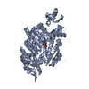 1lkxS S: Starting model for refinement |
|---|---|
| Similar structure data |


-Links
 PDBj
PDBj


- Assembly
| Deposited unit | 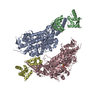
| ||||||||
|---|---|---|---|---|---|---|---|---|---|
| 1 | 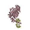
| ||||||||
| 2 | 
| ||||||||
| Unit cell |
|
-Components
| #1: Protein | Mass: 83939.688 Da / Num. of mol.: 2 / Fragment: MOTOR DOMAIN, RESIDUES 36-760 Source method: isolated from a genetically manipulated source Source: (gene. exp.) HOMO SAPIENS (human) / Plasmid: PFASTBAC DUAL / Cell line (production host): SF9 / Production host:   SPODOPTERA FRUGIPERDA (fall armyworm) / References: UniProt: O00159, EC: 3.6.4.1 SPODOPTERA FRUGIPERDA (fall armyworm) / References: UniProt: O00159, EC: 3.6.4.1#2: Protein | Mass: 16852.545 Da / Num. of mol.: 2 Source method: isolated from a genetically manipulated source Source: (gene. exp.) HOMO SAPIENS (human) / Plasmid: PFASTBAC DUAL / Cell line (production host): SF9 / Production host: SPODOPTERA FRUGIPERDA (fall armyworm) / References: UniProt: P62158, UniProt: P0DP23*PLUS#3: Chemical |   Mass: 24.305 Da / Num. of mol.: 3 / Source method: obtained synthetically / Formula: Mg Mass: 24.305 Da / Num. of mol.: 3 / Source method: obtained synthetically / Formula: Mg#4: Chemical |   Mass: 544.156 Da / Num. of mol.: 2 / Source method: obtained synthetically / Formula: C10H17N5O14P2V / Comment: energy-carrying molecule analogue*YM Mass: 544.156 Da / Num. of mol.: 2 / Source method: obtained synthetically / Formula: C10H17N5O14P2V / Comment: energy-carrying molecule analogue*YM#5: Water | ChemComp-HOH / |  Mass: 18.015 Da / Num. of mol.: 194 / Source method: isolated from a natural source / Formula: H2O Mass: 18.015 Da / Num. of mol.: 194 / Source method: isolated from a natural source / Formula: H2O |
|---|
-Experimental details
-Experiment
| Experiment | Method: X-RAY DIFFRACTION / Number of used crystals: 1 |
|---|
- Sample preparation
| Crystal | Density Matthews: 2.68 Å3/Da / Density % sol: 51.2 % / Description: NONE |
|---|---|
| Crystal grow | Details: 18% PEG3350, 0.2 M SODIUM MALONATE PH 7.0 |
-Data collection
| Diffraction | Mean temperature: 100 K |
|---|---|
| Diffraction source | Source: SYNCHROTRON / Site: ESRF  / Beamline: ID23-1 / Wavelength: 1.0645 / Beamline: ID23-1 / Wavelength: 1.0645 |
| Detector | Type: DECTRIS PILATUS 6M / Detector: PIXEL / Date: May 11, 2013 |
| Radiation | Monochromator: SILICON (1 1 1) CHANNEL-CUT / Protocol: SINGLE WAVELENGTH / Monochromatic (M) / Laue (L): M / Scattering type: x-ray |
| Radiation wavelength | Wavelength: 1.0645 Å / Relative weight: 1 |
| Reflection | Resolution: 2.74→47.94 Å / Num. obs: 51985 / % possible obs: 94 % / Observed criterion σ(I): 2 / Redundancy: 2.4 % / Biso Wilson estimate: 62.9 Å2 / Rmerge(I) obs: 0.07 / Net I/σ(I): 12.4 |
| Reflection shell | Resolution: 2.74→2.84 Å / Redundancy: 1.6 % / Rmerge(I) obs: 0.85 / Mean I/σ(I) obs: 2.15 / % possible all: 89 |
- Processing
| Software |
| ||||||||||||||||||
|---|---|---|---|---|---|---|---|---|---|---|---|---|---|---|---|---|---|---|---|
| Refinement | Method to determine structure: MOLECULAR REPLACEMENT Starting model: PDB ENTRY 1LKX Resolution: 2.74→47.94 Å / σ(F): 2 / Stereochemistry target values: ML
| ||||||||||||||||||
| Solvent computation | Shrinkage radii: 2 Å / VDW probe radii: 2 Å | ||||||||||||||||||
| Displacement parameters | Biso mean: 58.8 Å2 | ||||||||||||||||||
| Refinement step | Cycle: LAST / Resolution: 2.74→47.94 Å
| ||||||||||||||||||
| Refine LS restraints |
|
