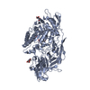
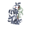
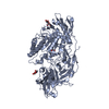
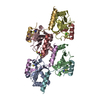
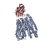
SEQUENCE AUTHOR STATES THE PUBLISHED SEQUENCE FOR DISTY MYOE IS INCORRECT IN A NUMBER OF PLACES. ...SEQUENCE AUTHOR STATES THE PUBLISHED SEQUENCE FOR DISTY MYOE IS INCORRECT IN A NUMBER OF PLACES. THE AUTHORS BELIEVE THEIR SEQUENCE TO BE MORE ACCURATE. THEY HAVE IDENTIFIED THE FOLLOWING CHANGES IN THEIR SEQUENCE/STRUCTURE FROM THE PUBLISHED ONE. THE DERIVED SEQUENCE OF MYOE WAS DIFFERENT FROM THE PUBLISHED SEQUENCE (URRUTIA ET AL., 1993, ACC. NR. L06805) IN 25 PLACES: WHILE SOME CHANGES CONCERNED NON-CONSERVED RESIDUES, ESPECIALLY IN THE REGIONS WHERE ADDITIONAL RESIDUES OR DELETIONS WERE FOUND, THE DERIVED SEQUENCE WAS IN BETTER AGREEMENT WITH THE AMINO ACID ALIGNMENT OF THE DICTYOSTELIUM MYOSINS. IN ONE REGION, A TEN RESIDUE SURFACE LOOP WAS FOUND TO BE COMPLETELY DIFFERENT. IN DETAIL, THE SEQUENCE CONTAINED THE FOLLOWING MODIFICATIONS COMPARED TO THE PUBLISHED ONE: D26E, R48T, I77M, I137L, R138D, F139 ABSENT, 140 ABSENT, N215D, L371I, S372I, I373N, V374C, H375T, R376T, G378K, T379G, P380 inserted, V427 inserted, R428 INSERTED, K429E, N440 INSERTED, N498I, D604V, I681N, R683T. THE AUTHORS STATE THEIR SEQUENCE COMPLETELY AGREES WITH THE FRAGMENT (AA270-1003) OBTAINED FROM THE DICTY GENOME SEQUENCING PROJECT.
In the structure databanks used in Yorodumi, some data are registered as the other names, "COVID-19 virus" and "2019-nCoV". Here are the details of the virus and the list of structure data.
Jan 31, 2019. EMDB accession codes are about to change! (news from PDBe EMDB page)
EMDB accession codes are about to change! (news from PDBe EMDB page)
The allocation of 4 digits for EMDB accession codes will soon come to an end. Whilst these codes will remain in use, new EMDB accession codes will include an additional digit and will expand incrementally as the available range of codes is exhausted. The current 4-digit format prefixed with “EMD-” (i.e. EMD-XXXX) will advance to a 5-digit format (i.e. EMD-XXXXX), and so on. It is currently estimated that the 4-digit codes will be depleted around Spring 2019, at which point the 5-digit format will come into force.
The EM Navigator/Yorodumi systems omit the EMD- prefix.
Related info.:Q: What is EMD? / ID/Accession-code notation in Yorodumi/EM Navigator
Yorodumi is a browser for structure data from EMDB, PDB, SASBDB, etc.
This page is also the successor to EM Navigator detail page, and also detail information page/front-end page for Omokage search.
The word "yorodu" (or yorozu) is an old Japanese word meaning "ten thousand". "mi" (miru) is to see.
Related info.:EMDB / PDB / SASBDB / Comparison of 3 databanks / Yorodumi Search / Aug 31, 2016. New EM Navigator & Yorodumi / Yorodumi Papers / Jmol/JSmol / Function and homology information / Changes in new EM Navigator and Yorodumi
 Movie
Movie Controller


 Open data
 Basic information
 Components
 Keywords
 Function and homology information
 X-RAY DIFFRACTION /
  Authors
 Citation
 Structure visualization
 Downloads & links
 Other downloads
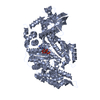

 PDBj
PDBj


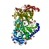
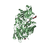
 Assembly


 Mass: 24.305 Da / Num. of mol.: 4 / Source method: obtained synthetically / Formula: Mg
 Mass: 114.939 Da / Num. of mol.: 4 / Source method: obtained synthetically / Formula: VO4
 Mass: 427.201 Da / Num. of mol.: 4 / Source method: obtained synthetically / Formula: C10H15N5O10P2 / Comment: ADP, energy-carrying molecule*YM
 Mass: 18.015 Da / Num. of mol.: 64 / Source method: isolated from a natural source / Formula: H2O
 Sample preparation

 Processing