 Movie
Movie Controller

[English] 日本語
 Yorodumi
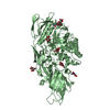
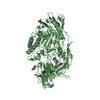
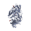
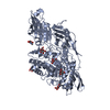
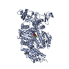
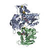
- PDB-6ik8: Crystal structure of tomato beta-galactosidase (TBG) 4 in complex... -
+ Open data

- Basic information
| Entry | Database: PDB / ID: 6ik8 | |||||||||
|---|---|---|---|---|---|---|---|---|---|---|
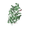
| Title | Crystal structure of tomato beta-galactosidase (TBG) 4 in complex with beta-1,6-galactobiose | |||||||||
 Components Components | Beta-galactosidase | |||||||||
 Keywords Keywords | HYDROLASE / Glycoside Hydrolase / Plant cell wall related enzyme / Fruit ripening | |||||||||
| Function / homology |  Function and homology information Function and homology informationbeta-galactosidase / beta-galactosidase activity / carbohydrate metabolic process Similarity search - Function | |||||||||
| Biological species |  | |||||||||
| Method |  X-RAY DIFFRACTION / SYNCHROTRON / MOLECULAR REPLACEMENT / Resolution: 2.8 Å X-RAY DIFFRACTION / SYNCHROTRON / MOLECULAR REPLACEMENT / Resolution: 2.8 Å | |||||||||
 Authors Authors | Matsuyama, K. / Nakae, S. / Igarashi, K. / Tada, T. / Ishimaru, M. | |||||||||
 Citation Citation | Journal: Planta / Year: 2020 Title: Substrate-recognition mechanism of tomato beta-galactosidase 4 using X-ray crystallography and docking simulation. Authors: Matsuyama, K. / Kondo, T. / Igarashi, K. / Sakamoto, T. / Ishimaru, M. | |||||||||
| History |
|
- Structure visualization
| Structure viewer | Molecule: MolmilJmol/JSmol |
|---|
- Downloads & links
-Download
| PDBx/mmCIF format | 6ik8.cif.gz | 295.1 KB | Display | PDBx/mmCIF format |
|---|---|---|---|---|
| PDB format | pdb6ik8.ent.gz | 235.7 KB | Display | PDB format |
| PDBx/mmJSON format | 6ik8.json.gz | Tree view | PDBx/mmJSON format | |
| Others |  Other downloads Other downloads |
-Validation report
| Arichive directory | https://data.pdbj.org/pub/pdb/validation_reports/ik/6ik8ftp://data.pdbj.org/pub/pdb/validation_reports/ik/6ik8 | HTTPS FTP |
|---|
-Related structure data
| Related structure data |  6ik5C  6ik6C  6ik7C  3w5gS S: Starting model for refinement C: citing same article ( |
|---|---|
| Similar structure data |

-Links
 PDBj
PDBj


- Assembly
| Deposited unit | 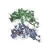
| ||||||||||||||||||
|---|---|---|---|---|---|---|---|---|---|---|---|---|---|---|---|---|---|---|---|
| 1 | 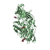
| ||||||||||||||||||
| 2 | 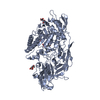
| ||||||||||||||||||
| Unit cell |
| ||||||||||||||||||
| Noncrystallographic symmetry (NCS) | NCS domain:
NCS domain segments: Component-ID: _ / Ens-ID: 1 / Beg auth comp-ID: GLU / Beg label comp-ID: GLU / End auth comp-ID: ALA / End label comp-ID: ALA / Refine code: _ / Auth seq-ID: 22 - 726 / Label seq-ID: 5 - 709
|
-Components
-Protein / Non-polymers , 2 types, 256 molecules BA
| #1: Protein | Mass: 79726.914 Da / Num. of mol.: 2 / Mutation: E181A Source method: isolated from a genetically manipulated source Source: (gene. exp.)  Komagataella pastoris (fungus) / Strain (production host): SMD1168H / References: UniProt: O81100, beta-galactosidase Komagataella pastoris (fungus) / Strain (production host): SMD1168H / References: UniProt: O81100, beta-galactosidase#6: Water | ChemComp-HOH / | Mass: 18.015 Da / Num. of mol.: 254 / Source method: isolated from a natural source / Formula: H2O |
|---|
-Sugars , 4 types, 7 molecules 

| #2: Polysaccharide | | #3: Polysaccharide | Source method: isolated from a genetically manipulated source #4: Sugar |  Type: D-saccharide, beta linking / Mass: 221.208 Da / Num. of mol.: 2 / Source method: isolated from a natural source / Formula: C8H15NO6 Type: D-saccharide, beta linking / Mass: 221.208 Da / Num. of mol.: 2 / Source method: isolated from a natural source / Formula: C8H15NO6#5: Sugar | ChemComp-GAL / |  Type: D-saccharide, beta linking / Mass: 180.156 Da / Num. of mol.: 1 Type: D-saccharide, beta linking / Mass: 180.156 Da / Num. of mol.: 1Source method: isolated from a genetically manipulated source Formula: C6H12O6 |
|---|
-Details
| Has protein modification | Y |
|---|
-Experimental details
-Experiment
| Experiment | Method: X-RAY DIFFRACTION / Number of used crystals: 1 |
|---|
- Sample preparation
| Crystal | Density Matthews: 2.22 Å3/Da / Density % sol: 44.66 % |
|---|---|
| Crystal grow | Temperature: 277 K / Method: vapor diffusion, sitting drop / pH: 7.3 / Details: 16% (w/v) PEG10000, 0.1M HEPES |
-Data collection
| Diffraction | Mean temperature: 100 K / Serial crystal experiment: N |
|---|---|
| Diffraction source | Source: SYNCHROTRON / Site: SPring-8  / Beamline: BL38B1 / Wavelength: 1 Å / Beamline: BL38B1 / Wavelength: 1 Å |
| Detector | Type: ADSC QUANTUM 315 / Detector: CCD / Date: Nov 1, 2016 |
| Radiation | Protocol: SINGLE WAVELENGTH / Monochromatic (M) / Laue (L): M / Scattering type: x-ray |
| Radiation wavelength | Wavelength: 1 Å / Relative weight: 1 |
| Reflection | Resolution: 2.8→50 Å / Num. obs: 35773 / % possible obs: 100 % / Redundancy: 6.6 % / Rmerge(I) obs: 0.152 / Net I/σ(I): 12.413 |
| Reflection shell | Resolution: 2.8→2.85 Å / Rmerge(I) obs: 0.755 / Mean I/σ(I) obs: 3 / Num. unique obs: 1744 / % possible all: 100 |
- Processing
| Software |
| ||||||||||||||||||||||||||||||||||||||||||||||||||||||||||||||||||||||||||||||||||||||||||||||||||||||||||||||||||||||||||||||||||||||||||||||||||||||||||||||||||||||||||||||||||||||
|---|---|---|---|---|---|---|---|---|---|---|---|---|---|---|---|---|---|---|---|---|---|---|---|---|---|---|---|---|---|---|---|---|---|---|---|---|---|---|---|---|---|---|---|---|---|---|---|---|---|---|---|---|---|---|---|---|---|---|---|---|---|---|---|---|---|---|---|---|---|---|---|---|---|---|---|---|---|---|---|---|---|---|---|---|---|---|---|---|---|---|---|---|---|---|---|---|---|---|---|---|---|---|---|---|---|---|---|---|---|---|---|---|---|---|---|---|---|---|---|---|---|---|---|---|---|---|---|---|---|---|---|---|---|---|---|---|---|---|---|---|---|---|---|---|---|---|---|---|---|---|---|---|---|---|---|---|---|---|---|---|---|---|---|---|---|---|---|---|---|---|---|---|---|---|---|---|---|---|---|---|---|---|---|
| Refinement | Method to determine structure: MOLECULAR REPLACEMENT Starting model: 3W5G Resolution: 2.8→48.02 Å / Cor.coef. Fo:Fc: 0.926 / Cor.coef. Fo:Fc free: 0.906 / SU B: 16.503 / SU ML: 0.312 / Cross valid method: THROUGHOUT / ESU R Free: 0.411 / Stereochemistry target values: MAXIMUM LIKELIHOOD
| ||||||||||||||||||||||||||||||||||||||||||||||||||||||||||||||||||||||||||||||||||||||||||||||||||||||||||||||||||||||||||||||||||||||||||||||||||||||||||||||||||||||||||||||||||||||
| Solvent computation | Ion probe radii: 0.8 Å / Shrinkage radii: 0.8 Å / VDW probe radii: 1.2 Å / Solvent model: MASK | ||||||||||||||||||||||||||||||||||||||||||||||||||||||||||||||||||||||||||||||||||||||||||||||||||||||||||||||||||||||||||||||||||||||||||||||||||||||||||||||||||||||||||||||||||||||
| Displacement parameters | Biso mean: 52.378 Å2
| ||||||||||||||||||||||||||||||||||||||||||||||||||||||||||||||||||||||||||||||||||||||||||||||||||||||||||||||||||||||||||||||||||||||||||||||||||||||||||||||||||||||||||||||||||||||
| Refinement step | Cycle: 1 / Resolution: 2.8→48.02 Å
| ||||||||||||||||||||||||||||||||||||||||||||||||||||||||||||||||||||||||||||||||||||||||||||||||||||||||||||||||||||||||||||||||||||||||||||||||||||||||||||||||||||||||||||||||||||||
| Refine LS restraints |
|