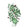
 Movie
Movie Controller


+ Open data

- Basic information
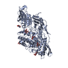
| Entry | Database: PDB / ID: 1br2 | ||||||
|---|---|---|---|---|---|---|---|
| Title | SMOOTH MUSCLE MYOSIN MOTOR DOMAIN COMPLEXED WITH MGADP.ALF4 | ||||||
 Components Components | MYOSIN | ||||||
 Keywords Keywords | MUSCLE PROTEIN | ||||||
| Function / homology |  Function and homology information Function and homology informationelastic fiber assembly / skeletal muscle myosin thick filament assembly / myofibril assembly / myosin light chain binding / myosin II binding / muscle myosin complex / myosin filament / actomyosin structure organization / cardiac muscle cell development / myosin II complex ...elastic fiber assembly / skeletal muscle myosin thick filament assembly / myofibril assembly / myosin light chain binding / myosin II binding / muscle myosin complex / myosin filament / actomyosin structure organization / cardiac muscle cell development / myosin II complex / structural constituent of muscle / smooth muscle contraction / microfilament motor activity / myofibril / ADP binding / actin filament binding / actin binding / calmodulin binding / magnesium ion binding / ATP binding / cytoplasm Similarity search - Function | ||||||
| Biological species |  | ||||||
| Method |  X-RAY DIFFRACTION / SYNCHROTRON / MOLECULAR REPLACEMENT / Resolution: 2.9 Å X-RAY DIFFRACTION / SYNCHROTRON / MOLECULAR REPLACEMENT / Resolution: 2.9 Å | ||||||
 Authors Authors | Dominguez, R. / Trybus, K.M. / Cohen, C. | ||||||
 Citation Citation | Journal: Cell(Cambridge,Mass.) / Year: 1998 Title: Crystal structure of a vertebrate smooth muscle myosin motor domain and its complex with the essential light chain: visualization of the pre-power stroke state. Authors: Dominguez, R. / Freyzon, Y. / Trybus, K.M. / Cohen, C. | ||||||
| History |
|
- Structure visualization
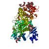
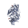
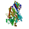
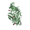
| Structure viewer | Molecule: MolmilJmol/JSmol |
|---|
- Downloads & links
-Download
| PDBx/mmCIF format | 1br2.cif.gz | 716.8 KB | Display | PDBx/mmCIF format |
|---|---|---|---|---|
| PDB format | pdb1br2.ent.gz | 590.1 KB | Display | PDB format |
| PDBx/mmJSON format | 1br2.json.gz | Tree view | PDBx/mmJSON format | |
| Others |  Other downloads Other downloads |
-Validation report
| Arichive directory | https://data.pdbj.org/pub/pdb/validation_reports/br/1br2ftp://data.pdbj.org/pub/pdb/validation_reports/br/1br2 | HTTPS FTP |
|---|
-Related structure data
| Related structure data | 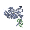 1br1C 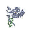 1br4C 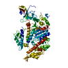 1mndS S: Starting model for refinement C: citing same article ( |
|---|---|
| Similar structure data |
-Links
 PDBj
PDBj


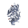
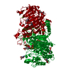
- Assembly
| Deposited unit | 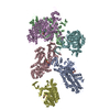
| ||||||||||||||||||||||||
|---|---|---|---|---|---|---|---|---|---|---|---|---|---|---|---|---|---|---|---|---|---|---|---|---|---|
| 1 | 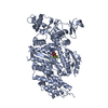
| ||||||||||||||||||||||||
| 2 | 
| ||||||||||||||||||||||||
| 3 | 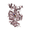
| ||||||||||||||||||||||||
| 4 | 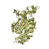
| ||||||||||||||||||||||||
| 5 | 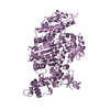
| ||||||||||||||||||||||||
| 6 | 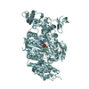
| ||||||||||||||||||||||||
| Unit cell |
| ||||||||||||||||||||||||
| Noncrystallographic symmetry (NCS) | NCS oper:
|
-Components
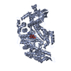
| #1: Protein | Mass: 90441.414 Da / Num. of mol.: 6 / Fragment: MOTOR DOMAIN Source method: isolated from a genetically manipulated source Details: LIGANDS MG, ADP, ALF(4) / Source: (gene. exp.)   Spodoptera frugiperda (fall armyworm) / References: UniProt: P10587, EC: 3.6.1.32 Spodoptera frugiperda (fall armyworm) / References: UniProt: P10587, EC: 3.6.1.32#2: Chemical | ChemComp-MG /   Mass: 24.305 Da / Num. of mol.: 6 / Source method: obtained synthetically / Formula: Mg Mass: 24.305 Da / Num. of mol.: 6 / Source method: obtained synthetically / Formula: Mg#3: Chemical | ChemComp-ALF /   Mass: 102.975 Da / Num. of mol.: 6 / Source method: obtained synthetically / Formula: AlF4 Mass: 102.975 Da / Num. of mol.: 6 / Source method: obtained synthetically / Formula: AlF4#4: Chemical | ChemComp-ADP /   Mass: 427.201 Da / Num. of mol.: 6 / Source method: obtained synthetically / Formula: C10H15N5O10P2 / Comment: ADP, energy-carrying molecule*YM Mass: 427.201 Da / Num. of mol.: 6 / Source method: obtained synthetically / Formula: C10H15N5O10P2 / Comment: ADP, energy-carrying molecule*YM#5: Water | ChemComp-HOH / |  Mass: 18.015 Da / Num. of mol.: 12 / Source method: isolated from a natural source / Formula: H2O Mass: 18.015 Da / Num. of mol.: 12 / Source method: isolated from a natural source / Formula: H2O |
|---|
-Experimental details
-Experiment
| Experiment | Method: X-RAY DIFFRACTION / Number of used crystals: 1 |
|---|
- Sample preparation
| Crystal | Density Matthews: 2.5 Å3/Da / Density % sol: 51 % | ||||||||||||||||||||||||||||||||||||||||||||||||
|---|---|---|---|---|---|---|---|---|---|---|---|---|---|---|---|---|---|---|---|---|---|---|---|---|---|---|---|---|---|---|---|---|---|---|---|---|---|---|---|---|---|---|---|---|---|---|---|---|---|
| Crystal grow | Temperature: 277 K / pH: 6.8 Details: 12% PEG8000 100MM IMIDAZOLE PH 6.8 150MM CACL2 2MM MGADP 2MM AL(NO3)3 8MM NAF PROTEIN CONCENTRATION: 10MG/ML TEMPERATURE: 4 DEGREES CENTIGRADE, temperature 277K | ||||||||||||||||||||||||||||||||||||||||||||||||
| Crystal grow | *PLUS Method: vapor diffusion, sitting drop | ||||||||||||||||||||||||||||||||||||||||||||||||
| Components of the solutions | *PLUS
|
-Data collection
| Diffraction | Mean temperature: 100 K |
|---|---|
| Diffraction source | Source: SYNCHROTRON / Site: CHESS  / Beamline: F1 / Wavelength: 0.918 / Beamline: F1 / Wavelength: 0.918 |
| Detector | Detector: CCD / Date: Jan 1, 1997 |
| Radiation | Monochromatic (M) / Laue (L): M / Scattering type: x-ray |
| Radiation wavelength | Wavelength: 0.918 Å / Relative weight: 1 |
| Reflection | Resolution: 2.9→20 Å / Num. obs: 99071 / % possible obs: 83.7 % / Redundancy: 3.3 % / Rsym value: 0.072 / Net I/σ(I): 28 |
| Reflection shell | Resolution: 2.9→3.12 Å / Redundancy: 3 % / Mean I/σ(I) obs: 14.5 / Rsym value: 0.215 / % possible all: 67.7 |
| Reflection | *PLUS Num. measured all: 326933 / Rmerge(I) obs: 0.072 |
| Reflection shell | *PLUS % possible obs: 67.7 % / Rmerge(I) obs: 0.215 |
- Processing
| Software |
| ||||||||||||||||||||||||||||||||||||||||||||||||||||||||||||
|---|---|---|---|---|---|---|---|---|---|---|---|---|---|---|---|---|---|---|---|---|---|---|---|---|---|---|---|---|---|---|---|---|---|---|---|---|---|---|---|---|---|---|---|---|---|---|---|---|---|---|---|---|---|---|---|---|---|---|---|---|---|
| Refinement | Method to determine structure: MOLECULAR REPLACEMENT Starting model: PDB ENTRY 1MND Resolution: 2.9→10 Å / Data cutoff high absF: 0 / Data cutoff low absF: 0 / Isotropic thermal model: RESTRAINED / Cross valid method: THROUGHOUT / σ(F): 0
| ||||||||||||||||||||||||||||||||||||||||||||||||||||||||||||
| Displacement parameters | Biso mean: 41.5 Å2 | ||||||||||||||||||||||||||||||||||||||||||||||||||||||||||||
| Refine analyze | Luzzati coordinate error free: 0.5 Å | ||||||||||||||||||||||||||||||||||||||||||||||||||||||||||||
| Refinement step | Cycle: LAST / Resolution: 2.9→10 Å
| ||||||||||||||||||||||||||||||||||||||||||||||||||||||||||||
| Refine LS restraints |
| ||||||||||||||||||||||||||||||||||||||||||||||||||||||||||||
| Refine LS restraints NCS | NCS model details: RESTRAINTS | ||||||||||||||||||||||||||||||||||||||||||||||||||||||||||||
| Xplor file |
| ||||||||||||||||||||||||||||||||||||||||||||||||||||||||||||
| Software | *PLUS Name: X-PLOR / Version: 3.851 / Classification: refinement | ||||||||||||||||||||||||||||||||||||||||||||||||||||||||||||
| Refinement | *PLUS | ||||||||||||||||||||||||||||||||||||||||||||||||||||||||||||
| Solvent computation | *PLUS | ||||||||||||||||||||||||||||||||||||||||||||||||||||||||||||
| Displacement parameters | *PLUS | ||||||||||||||||||||||||||||||||||||||||||||||||||||||||||||
| Refine LS restraints | *PLUS
|
