 Movie
Movie Controller


+ Open data

- Basic information
| Entry | Database: PDB / ID: 1fgt | ||||||
|---|---|---|---|---|---|---|---|
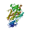
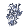
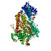
| Title | LIPOXYGENASE-1 (SOYBEAN) AT 100K, Q697N MUTANT | ||||||
 Components Components | SEED LIPOXYGENASE-1 | ||||||
 Keywords Keywords | OXIDOREDUCTASE / DIOXYGENASE / LIPOXYGENASE / METALLOPROTEIN / FATTY ACIDS | ||||||
| Function / homology |  Function and homology information Function and homology informationlinolenate 9R-lipoxygenase activity / linoleate 13S-lipoxygenase / linoleate 13S-lipoxygenase activity / oxylipin biosynthetic process / lipid oxidation / oxidoreductase activity, acting on single donors with incorporation of molecular oxygen, incorporation of two atoms of oxygen / fatty acid oxidation / fatty acid biosynthetic process / iron ion binding / cytoplasm Similarity search - Function | ||||||
| Biological species |  | ||||||
| Method |  X-RAY DIFFRACTION / SYNCHROTRON / IR / Resolution: 1.62 Å X-RAY DIFFRACTION / SYNCHROTRON / IR / Resolution: 1.62 Å | ||||||
 Authors Authors | Tomchick, D.R. / Minor, W. / Holman, T. | ||||||
 Citation Citation | Journal: Biochemistry / Year: 2001 Title: Structural and functional characterization of second-coordination sphere mutants of soybean lipoxygenase-1. Authors: Tomchick, D.R. / Phan, P. / Cymborowski, M. / Minor, W. / Holman, T.R. #1: Journal: Biochemistry / Year: 1996Title: CRYSTAL STRUCTURE OF SOYBEAN LIPOXYGENASE L-1 AT 1.4 A RESOLUTION Authors: MINOR, W. / STECZKO, J. / STEC, B. / OTWINOWSKI, Z. / BOLIN, J.T. / WALTER, R. / AXELROD, B. #2: Journal: CURR.OPIN.STRUCT.BIOL. / Year: 1994Title: THE STRUCTURE AND FUNCTION OF LIPOXYGENASE Authors: NELSON, M.J. / SEITZ, S.P. #3: Journal: J.Am.Chem.Soc. / Year: 1996Title: EXPERIMENTAL EVIDENCE FOR EXTENSIVE TUNNELING OF HYDROGEN IN THE LIPOXYGENASE REACTION: IMPLICATIONS FOR ENZYME CATALYSIS Authors: JONSSON, T. / GLICKMAN, M.H. / SUN, S. / KLINMAN, J.P. #4: Journal: Biochemistry / Year: 1995Title: NATURE OF THE RATE-LIMITING STEPS IN THE SOYBEAN LIPOXYGENASE-1 REACTION Authors: GLICKMAN, M.H. / KLINMAN, J.P. #5: Journal: Science / Year: 1993Title: THE THREE-DIMENSIONAL STRUCTURE OF AN ARACHIDONIC ACID 15-LIPOXYGENASE Authors: BOYINGTON, J.C. / GAFFNEY, B.J. / AMZEL, L.M. #6: Journal: Biochemistry / Year: 1993Title: CRYSTALLOGRAPHIC DETERMINATION OF THE ACTIVE SITE IRON AND ITS LIGANDS IN SOYBEAN LIPOXYGENASE L-1 Authors: MINOR, W. / STECZKO, J. / BOLIN, J.T. / OTWINOWSKI, Z. / AXELROD, B. #7: Journal: J.Biol.Chem. / Year: 1990Title: CRYSTALLIZATION AND PRELIMINARY X-RAY INVESTIGATION OF LIPOXYGENASE 1 FROM SOYBEANS Authors: STECZKO, J. / MUCHMORE, C.R. / L SMITH, J. / AXELROD, B. | ||||||
| History |
|
- Structure visualization
| Structure viewer | Molecule: MolmilJmol/JSmol |
|---|
- Downloads & links
-Download
| PDBx/mmCIF format | 1fgt.cif.gz | 193.5 KB | Display | PDBx/mmCIF format |
|---|---|---|---|---|
| PDB format | pdb1fgt.ent.gz | 150.2 KB | Display | PDB format |
| PDBx/mmJSON format | 1fgt.json.gz | Tree view | PDBx/mmJSON format | |
| Others |  Other downloads Other downloads |
-Validation report
| Arichive directory | https://data.pdbj.org/pub/pdb/validation_reports/fg/1fgtftp://data.pdbj.org/pub/pdb/validation_reports/fg/1fgt | HTTPS FTP |
|---|
-Related structure data
| Related structure data |  1f8nSC  1fgmC 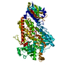 1fgoC  1fgqC  1fgrC S: Starting model for refinement C: citing same article ( |
|---|---|
| Similar structure data |


-Links
 PDBj
PDBj- Assembly
| Deposited unit | 
| ||||||||
|---|---|---|---|---|---|---|---|---|---|
| 1 |
| ||||||||
| Unit cell |
|
-Components
| #1: Protein | Mass: 94466.102 Da / Num. of mol.: 1 / Mutation: Q697N Source method: isolated from a genetically manipulated source Source: (gene. exp.)  |
|---|---|
| #2: Chemical | ChemComp-FE /   Mass: 55.845 Da / Num. of mol.: 1 / Source method: obtained synthetically / Formula: Fe Mass: 55.845 Da / Num. of mol.: 1 / Source method: obtained synthetically / Formula: Fe |
| #3: Water | ChemComp-HOH /  Mass: 18.015 Da / Num. of mol.: 753 / Source method: isolated from a natural source / Formula: H2O Mass: 18.015 Da / Num. of mol.: 753 / Source method: isolated from a natural source / Formula: H2O |
-Experimental details
-Experiment
| Experiment | Method: X-RAY DIFFRACTION / Number of used crystals: 1 |
|---|
- Sample preparation
| Crystal | Density Matthews: 2.1 Å3/Da / Density % sol: 41.5 % | ||||||||||||||||||||||||||||||
|---|---|---|---|---|---|---|---|---|---|---|---|---|---|---|---|---|---|---|---|---|---|---|---|---|---|---|---|---|---|---|---|
| Crystal grow | Temperature: 293 K / Method: vapor diffusion, sitting drop / pH: 5.5 Details: PEG 3350, SODIUM ACETATE, pH 5.5, VAPOR DIFFUSION, SITTING DROP, temperature 293K | ||||||||||||||||||||||||||||||
| Crystal grow | *PLUS Temperature: 21 ℃ / pH: 5.6 / Details: Minor, W., (1996) Biochemistry, 35, 10687. | ||||||||||||||||||||||||||||||
| Components of the solutions | *PLUS
|
-Data collection
| Diffraction | Mean temperature: 100 K |
|---|---|
| Diffraction source | Source: SYNCHROTRON / Site: APS  / Beamline: 19-ID / Wavelength: 0.97918 / Beamline: 19-ID / Wavelength: 0.97918 |
| Detector | Type: SBC-2 / Detector: CCD / Date: Dec 1, 1999 |
| Radiation | Protocol: SINGLE WAVELENGTH / Monochromatic (M) / Laue (L): M / Scattering type: x-ray |
| Radiation wavelength | Wavelength: 0.97918 Å / Relative weight: 1 |
| Reflection | Resolution: 1.62→20 Å / Num. all: 107678 / Num. obs: 104391 / % possible obs: 96.9 % / Observed criterion σ(F): 0 / Observed criterion σ(I): 0 / Redundancy: 4.7 % / Biso Wilson estimate: 27.6 Å2 / Rmerge(I) obs: 0.043 / Net I/σ(I): 25.2 |
| Reflection shell | Resolution: 1.62→1.68 Å / Redundancy: 4 % / Rmerge(I) obs: 0.337 / Num. unique all: 9958 / % possible all: 92.7 |
| Reflection | *PLUS Num. measured all: 490154 |
- Processing
| Software |
| ||||||||||||||||||||||||||||||||||||
|---|---|---|---|---|---|---|---|---|---|---|---|---|---|---|---|---|---|---|---|---|---|---|---|---|---|---|---|---|---|---|---|---|---|---|---|---|---|
| Refinement | Method to determine structure: IR Starting model: 1F8N Resolution: 1.62→20 Å / σ(F): 0 / σ(I): 0 / Stereochemistry target values: Engh & Huber
| ||||||||||||||||||||||||||||||||||||
| Solvent computation | Solvent model: anisotropic / Bsol: 46.318 Å2 / ksol: 0.3509 e/Å3 | ||||||||||||||||||||||||||||||||||||
| Displacement parameters | Biso mean: 24.15 Å2
| ||||||||||||||||||||||||||||||||||||
| Refine analyze |
| ||||||||||||||||||||||||||||||||||||
| Refinement step | Cycle: LAST / Resolution: 1.62→20 Å
| ||||||||||||||||||||||||||||||||||||
| Refine LS restraints |
| ||||||||||||||||||||||||||||||||||||
| LS refinement shell | Resolution: 1.62→1.68 Å / Total num. of bins used: 10
| ||||||||||||||||||||||||||||||||||||
| Software | *PLUS Name: CNS / Classification: refinement | ||||||||||||||||||||||||||||||||||||
| Refinement | *PLUS Lowest resolution: 20 Å / σ(F): 0 / % reflection Rfree: 4.5 % / Rfactor obs: 0.183 | ||||||||||||||||||||||||||||||||||||
| Solvent computation | *PLUS | ||||||||||||||||||||||||||||||||||||
| Displacement parameters | *PLUS | ||||||||||||||||||||||||||||||||||||
| Refine LS restraints | *PLUS
|
