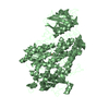
 Movie
Movie Controller


+ Open data

- Basic information
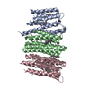
| Entry | Database: PDB / ID: 1lnh | ||||||
|---|---|---|---|---|---|---|---|
| Title | LIPOXYGENASE-3(SOYBEAN) NON-HEME FE(II) METALLOPROTEIN | ||||||
 Components Components | LIPOXYGENASE-3 | ||||||
 Keywords Keywords | OXIDOREDUCTASE / METALLOPROTEIN / FE(II) COMPLEX | ||||||
| Function / homology |  Function and homology information Function and homology informationlinoleate 9S-lipoxygenase / linoleate 9S-lipoxygenase activity / oxylipin biosynthetic process / lipid oxidation / oxidoreductase activity, acting on single donors with incorporation of molecular oxygen, incorporation of two atoms of oxygen / fatty acid biosynthetic process / iron ion binding / cytoplasm Similarity search - Function | ||||||
| Biological species |  | ||||||
| Method |  X-RAY DIFFRACTION / MOLECULAR REPLACEMENT / Resolution: 2.6 Å X-RAY DIFFRACTION / MOLECULAR REPLACEMENT / Resolution: 2.6 Å | ||||||
 Authors Authors | Skrzypczak-Jankun, E. | ||||||
 Citation Citation | Journal: Proteins / Year: 1997 Title: Structure of soybean lipoxygenase L3 and a comparison with its L1 isoenzyme. Authors: Skrzypczak-Jankun, E. / Amzel, L.M. / Kroa, B.A. / Funk Jr., M.O. #1: Journal: J.Mol.Struct. / Year: 1996Title: Lipoxygenase-A Molecular Complex with a Non-Heme Iron Authors: Skrzypczak-Jankun, E. / Funk Junior, M.O. / Boyington, J.C. / Amzel, L.M. #2: Journal: Biochemistry / Year: 1994Title: Position 713 is Critical for Catalysis But not Iron Binding in Soybean Lipoxygenase 3 Authors: Kramer, J.A. / Johnson, K.R. / Dunham, W.R. / Sands, R.H. / Funk Junior, M.O. #3: Journal: J.Mol.Biol. / Year: 1990Title: Crystallization and Preliminary X-Ray Characterization of a Soybean Seed Lipoxygenase Authors: Stallings, W.C. / Kroa, B.A. / Carroll, R.T. / Metzger, A.L. / Funk, M.O. | ||||||
| History |
|
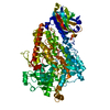
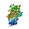
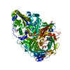
- Structure visualization
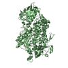
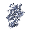
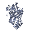
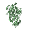
| Structure viewer | Molecule: MolmilJmol/JSmol |
|---|
- Downloads & links
-Download
| PDBx/mmCIF format | 1lnh.cif.gz | 183 KB | Display | PDBx/mmCIF format |
|---|---|---|---|---|
| PDB format | pdb1lnh.ent.gz | 142.8 KB | Display | PDB format |
| PDBx/mmJSON format | 1lnh.json.gz | Tree view | PDBx/mmJSON format | |
| Others |  Other downloads Other downloads |
-Validation report
| Arichive directory | https://data.pdbj.org/pub/pdb/validation_reports/ln/1lnhftp://data.pdbj.org/pub/pdb/validation_reports/ln/1lnh | HTTPS FTP |
|---|
-Related structure data


-Links
 PDBj
PDBj


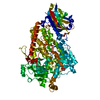
- Assembly
| Deposited unit | 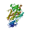
| ||||||||
|---|---|---|---|---|---|---|---|---|---|
| 1 |
| ||||||||
| Unit cell |
|
-Components
| #1: Protein | Mass: 96919.000 Da / Num. of mol.: 1 / Source method: isolated from a natural source / Source: (natural) |
|---|---|
| #2: Chemical | ChemComp-FE2 /   Mass: 55.845 Da / Num. of mol.: 1 / Source method: obtained synthetically / Formula: Fe Mass: 55.845 Da / Num. of mol.: 1 / Source method: obtained synthetically / Formula: Fe |
| #3: Water | ChemComp-HOH /  Mass: 18.015 Da / Num. of mol.: 170 / Source method: isolated from a natural source / Formula: H2O Mass: 18.015 Da / Num. of mol.: 170 / Source method: isolated from a natural source / Formula: H2O |
| Compound details | THE NUMBERING OF THE SECONDARY STRUCTURE ELEMENTS HAS SYMBOLS AFTER SOYBEAN LIPOXYGENASE L1 AS IN ...THE NUMBERING OF THE SECONDARY STRUCTURE ELEMENTS HAS SYMBOLS AFTER SOYBEAN LIPOXYGENA |
| Nonpolymer details | THE IRON BINDING SITE CONSISTS OF HIS 518, HIS 523, HIS 709 AND ILE 857(C-TERMINAL) THAT ARE ...THE IRON BINDING SITE CONSISTS OF HIS 518, HIS 523, HIS 709 AND ILE 857(C-TERMINAL) THAT ARE COVALENTLY |
-Experimental details
-Experiment
| Experiment | Method: X-RAY DIFFRACTION / Number of used crystals: 2 |
|---|
- Sample preparation
| Crystal | Density Matthews: 2.46 Å3/Da / Density % sol: 50 % | |||||||||||||||||||||||||||||||||||||||||||||||||||||||||||||||
|---|---|---|---|---|---|---|---|---|---|---|---|---|---|---|---|---|---|---|---|---|---|---|---|---|---|---|---|---|---|---|---|---|---|---|---|---|---|---|---|---|---|---|---|---|---|---|---|---|---|---|---|---|---|---|---|---|---|---|---|---|---|---|---|---|
| Crystal grow | pH: 5.3 / Details: pH 5.3 | |||||||||||||||||||||||||||||||||||||||||||||||||||||||||||||||
| Crystal grow | *PLUS Temperature: 23 ℃ / Method: vapor diffusion, sitting drop | |||||||||||||||||||||||||||||||||||||||||||||||||||||||||||||||
| Components of the solutions | *PLUS
|
-Data collection
| Diffraction | Mean temperature: 296 K |
|---|---|
| Diffraction source | Source: ROTATING ANODE / Wavelength: 1.5418 |
| Detector | Type: RIGAKU / Detector: IMAGE PLATE / Date: 1994 |
| Radiation | Monochromator: GRAPHITE(002) / Monochromatic (M) / Laue (L): M / Scattering type: x-ray |
| Radiation wavelength | Wavelength: 1.5418 Å / Relative weight: 1 |
| Reflection | Resolution: 2.6→61.6 Å / Num. obs: 23403 / % possible obs: 80.1 % / Observed criterion σ(I): 2 / Rmerge(I) obs: 0.062 |
| Reflection shell | Resolution: 2.6→2.75 Å / Rmerge(I) obs: 0.117 / Mean I/σ(I) obs: 3.39 / % possible all: 43.6 |
| Reflection | *PLUS Num. measured all: 74331 |
| Reflection shell | *PLUS % possible obs: 48 % |
- Processing
| Software |
| ||||||||||||||||||||||||||||||||||||||||||||||||||||||||||||
|---|---|---|---|---|---|---|---|---|---|---|---|---|---|---|---|---|---|---|---|---|---|---|---|---|---|---|---|---|---|---|---|---|---|---|---|---|---|---|---|---|---|---|---|---|---|---|---|---|---|---|---|---|---|---|---|---|---|---|---|---|---|
| Refinement | Method to determine structure: MOLECULAR REPLACEMENT Starting model: FOR MOLECULAR REPLACEMENT: 1SBL FOR SOYBEAN LIPOXYGENASE-1. THE MODEL WAS TRUNCATED TO INCLUDE ONLY THE ATOMS (7854) COMMON WITH LIPOXYGENASE-3 SEQUENCE (ENTRY 1SBL WAS REPLACED LATER ...Starting model: FOR MOLECULAR REPLACEMENT: 1SBL FOR SOYBEAN LIPOXYGENASE-1. THE MODEL WAS TRUNCATED TO INCLUDE ONLY THE ATOMS (7854) COMMON WITH LIPOXYGENASE-3 SEQUENCE (ENTRY 1SBL WAS REPLACED LATER IN PDB BY 2SBL WITH BETTER REFINED MODEL). Resolution: 2.6→8 Å / σ(F): 2
| ||||||||||||||||||||||||||||||||||||||||||||||||||||||||||||
| Displacement parameters | Biso mean: 31.7 Å2 | ||||||||||||||||||||||||||||||||||||||||||||||||||||||||||||
| Refine analyze | Luzzati coordinate error obs: 0.2 Å | ||||||||||||||||||||||||||||||||||||||||||||||||||||||||||||
| Refinement step | Cycle: LAST / Resolution: 2.6→8 Å
| ||||||||||||||||||||||||||||||||||||||||||||||||||||||||||||
| Refine LS restraints |
| ||||||||||||||||||||||||||||||||||||||||||||||||||||||||||||
| Software | *PLUS Name: X-PLOR / Version: 3.1 / Classification: refinement | ||||||||||||||||||||||||||||||||||||||||||||||||||||||||||||
| Refine LS restraints | *PLUS
|
