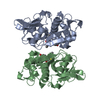
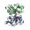
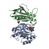
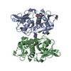
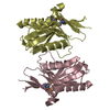
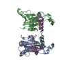
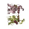
Entry Database : PDB / ID : 3h8dTitle Crystal structure of Myosin VI in complex with Dab2 peptide Disabled homolog 2 Myosin-VI Keywords / / / / / / / / / / / / / / / / / / / / / / / / / / / / Function / homology Function Domain/homology Component
/ / / / / / / / / / / / / / / / / / / / / / / / / / / / / / / / / / / / / / / / / / / / / / / / / / / / / / / / / / / / / / / / / / / / / / / / / / / / / / / / / / / / / / / / / / / / / / / / / / / / / / / / / / / / / / / / / / / / / / / / / / / / / / / / / / / / / / / / / / Biological species Mus musculus (house mouse)Rattus norvegicus (Norway rat)Method / / / Resolution : 2.2 Å Authors Yu, C. / Feng, W. / Wei, Z. / Zhang, M. Journal : Cell(Cambridge,Mass.) / Year : 2009Title : Myosin VI undergoes cargo-mediated dimerizationAuthors : Yu, C. / Feng, W. / Wei, Z. / Miyanoiri, Y. / Wen, W. / Zhao, Y. / Zhang, M. History Deposition Apr 29, 2009 Deposition site / Processing site Revision 1.0 Sep 29, 2009 Provider / Type Revision 1.1 Jul 13, 2011 Group Revision 1.2 Mar 20, 2024 Group / Database references / Derived calculationsCategory chem_comp_atom / chem_comp_bond ... chem_comp_atom / chem_comp_bond / database_2 / struct_ref_seq_dif / struct_site Item _database_2.pdbx_DOI / _database_2.pdbx_database_accession ... _database_2.pdbx_DOI / _database_2.pdbx_database_accession / _struct_ref_seq_dif.details / _struct_site.pdbx_auth_asym_id / _struct_site.pdbx_auth_comp_id / _struct_site.pdbx_auth_seq_id
Show all Show less
 Movie
Movie Controller


 Open data
 Basic information
 Components
 Keywords
 Function and homology information
 X-RAY DIFFRACTION /
  Authors
 Citation
 Structure visualization
 Downloads & links
 Other downloads
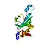


 PDBj
PDBj


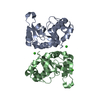
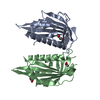
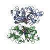
 Assembly
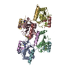


 Mass: 58.082 Da / Num. of mol.: 3 / Source method: obtained synthetically / Formula: CNS
 Mass: 154.251 Da / Num. of mol.: 1 / Source method: obtained synthetically / Formula: C4H10O2S2
 Mass: 35.453 Da / Num. of mol.: 1 / Source method: obtained synthetically / Formula: Cl
 Sample preparation
 Processing