[English] 日本語
Yorodumi
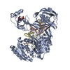
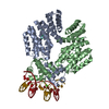
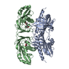
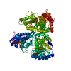
- PDB-3k90: The Abscisic acid receptor PYR1 in complex with Abscisic Acid -

+
Open data


ID or keywords:

Loading...

-
Basic information

Entry
Database: PDB / ID: 3k90
TitleThe Abscisic acid receptor PYR1 in complex with Abscisic Acid
ComponentsPutative uncharacterized protein
KeywordsHORMONE RECEPTOR / HYDROLASE REGULATOR / GENE REGULATOR / HORMONE / PLANT PROTEIN / SIGNALING PROTEIN / START domain / Bet V I domain / Hormone-Receptor complex / Regulator of protein phosphatase type 2C
Function / homology
Function and homology information


positive regulation of response to water deprivation / regulation of protein serine/threonine phosphatase activity / plant-type vacuole membrane / protein phosphatase inhibitor complex / abscisic acid binding / abscisic acid-activated signaling pathway / protein phosphatase inhibitor activity / ubiquitin-like protein ligase binding / signaling receptor activity / protein homodimerization activity ...positive regulation of response to water deprivation / regulation of protein serine/threonine phosphatase activity / plant-type vacuole membrane / protein phosphatase inhibitor complex / abscisic acid binding / abscisic acid-activated signaling pathway / protein phosphatase inhibitor activity / ubiquitin-like protein ligase binding / signaling receptor activity / protein homodimerization activity / identical protein binding / nucleus / plasma membrane / cytoplasm / cytosol
Similarity search - Function
Polyketide cyclase / dehydrase and lipid transport / Polyketide cyclase/dehydrase / Polyketide cyclase / dehydrase and lipid transport / START domain / Alpha-D-Glucose-1,6-Bisphosphate; Chain A, domain 4 / START-like domain superfamily / 2-Layer Sandwich / Alpha Beta
Similarity search - Domain/homology
Chem-A8S / ACETIC ACID / Abscisic acid receptor PYR1
Similarity search - Component
Biological speciesArabidopsis thaliana (thale cress)
MethodX-RAY DIFFRACTION / SYNCHROTRON / MOLECULAR REPLACEMENT / Resolution: 2 Å
AuthorsDupeux, F.D. / Santiago, J. / Rodriguez, P.L. / Marquez, J.A.
CitationJournal: Nature / Year: 2009
Title: The abscisic acid receptor PYR1 in complex with abscisic acid.
Authors: Santiago, J. / Dupeux, F. / Round, A. / Antoni, R. / Park, S.Y. / Jamin, M. / Cutler, S.R. / Rodriguez, P.L. / Marquez, J.A.
History
DepositionOct 15, 2009Deposition site: RCSB / Processing site: PDBJ
Revision 1.0Nov 10, 2009Provider: repository / Type: Initial release
Revision 1.1Jul 13, 2011Group: Non-polymer description / Version format compliance
Revision 1.2Nov 10, 2021Group: Database references / Derived calculations / Structure summary
Category: chem_comp / database_2 ...chem_comp / database_2 / struct_ref_seq_dif / struct_site
Item: _chem_comp.pdbx_synonyms / _database_2.pdbx_DOI ..._chem_comp.pdbx_synonyms / _database_2.pdbx_DOI / _database_2.pdbx_database_accession / _struct_ref_seq_dif.details / _struct_site.pdbx_auth_asym_id / _struct_site.pdbx_auth_comp_id / _struct_site.pdbx_auth_seq_id
Revision 1.3Nov 1, 2023Group: Data collection / Refinement description
Category: chem_comp_atom / chem_comp_bond / pdbx_initial_refinement_model

-
Structure visualization

Structure viewerMolecule:
MolmilJmol/JSmol

Downloads & links

-
Assembly

Deposited unit
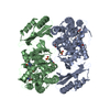
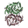
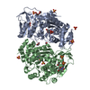
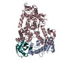
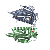
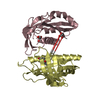
A: Putative uncharacterized protein
B: Putative uncharacterized protein
C: Putative uncharacterized protein
D: Putative uncharacterized protein
hetero molecules


Theoretical massNumber of molelcules
Total (without water)87,68610
Polymers86,8214
Non-polymers8656
Water9,908550
1


  • Idetical with deposited unit
  • defined by software
TypeNameSymmetry operationNumber
identity operation1_555x,y,z1
Buried area6600 Å2
ΔGint-44 kcal/mol
Surface area32930 Å2
MethodPISA
2
A: Putative uncharacterized protein
B: Putative uncharacterized protein
hetero molecules


Theoretical massNumber of molelcules
Total (without water)43,8275
Polymers43,4112
Non-polymers4163
Water362
TypeNameSymmetry operationNumber
identity operation1_555x,y,z1
Buried area1650 Å2
ΔGint-14 kcal/mol
Surface area17800 Å2
MethodPISA
3
C: Putative uncharacterized protein
D: Putative uncharacterized protein
hetero molecules


Theoretical massNumber of molelcules
Total (without water)43,8595
Polymers43,4112
Non-polymers4493
Water362
TypeNameSymmetry operationNumber
identity operation1_555x,y,z1
Buried area2020 Å2
ΔGint-16 kcal/mol
Surface area18050 Å2
MethodPISA
Unit cell
Length a, b, c (Å)49.810, 61.280, 72.550
Angle α, β, γ (deg.)105.65, 102.23, 89.91
Int Tables number1
Space group name H-MP1

-
Components

#1: Protein
Putative uncharacterized protein / PYR1 / Putative uncharacterized protein AT4g17870 / Putative uncharacterized protein T6K21.50


Mass: 21705.340 Da / Num. of mol.: 4 / Mutation: P2A
Source method: isolated from a genetically manipulated source
Source: (gene. exp.) Arabidopsis thaliana (thale cress) / Gene: AT4g17870, T6K21.50 / Production host: Escherichia coli (E. coli) / Strain (production host): BL21 DE3 / References: UniProt: O49686
#2: Chemical ChemComp-A8S / (2Z,4E)-5-[(1S)-1-hydroxy-2,6,6-trimethyl-4-oxocyclohex-2-en-1-yl]-3-methylpenta-2,4-dienoic acid / (+)-abscisic acid / (2Z,4E)-5-[(1S)-1-hydroxy-2,6,6-trimethyl-4-oxo-2-cyclohexen-1-yl]-3-methyl-2,4-pentadienoic acid


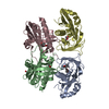
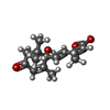
Mass: 264.317 Da / Num. of mol.: 2 / Source method: obtained synthetically / Formula: C15H20O4 / Comment: hormone*YM
#3: Chemical ChemComp-GOL / GLYCEROL / GLYCERIN / PROPANE-1,2,3-TRIOL


Mass: 92.094 Da / Num. of mol.: 3 / Source method: obtained synthetically / Formula: C3H8O3
#4: Chemical ChemComp-ACY / ACETIC ACID


Mass: 60.052 Da / Num. of mol.: 1 / Source method: obtained synthetically / Formula: C2H4O2
#5: Water ChemComp-HOH / water


Mass: 18.015 Da / Num. of mol.: 550 / Source method: isolated from a natural source / Formula: H2O

-
Experimental details

-
Experiment

ExperimentMethod: X-RAY DIFFRACTION / Number of used crystals: 1

-
Sample preparation

CrystalDensity Matthews: 2.4 Å3/Da / Density % sol: 48.67 %
Crystal growTemperature: 298 K / Method: vapor diffusion, hanging drop / pH: 6.5
Details: 0.2 M sodium magnesium acetate, 0.1M sodium cacodylate pH 6.5, 18% PEG 8000, VAPOR DIFFUSION, HANGING DROP, temperature 298K

-
Data collection

DiffractionMean temperature: 100 K
Diffraction sourceSource: SYNCHROTRON / Site: ESRF / Beamline: ID14-4 / Wavelength: 0.933 Å
DetectorType: ADSC QUANTUM 315r / Detector: CCD / Date: May 6, 2009 / Details: mirrors
RadiationMonochromator: channel cut ESRF monochromator / Protocol: SINGLE WAVELENGTH / Monochromatic (M) / Laue (L): M / Scattering type: x-ray
Radiation wavelengthWavelength: 0.933 Å / Relative weight: 1
ReflectionResolution: 2→25 Å / Num. all: 54557 / Num. obs: 50927 / % possible obs: 93.3 % / Observed criterion σ(F): 0 / Observed criterion σ(I): 0
Reflection shellResolution: 2→2.1 Å / % possible all: 86.9

-
Processing

Software
NameVersionClassification
XDSdata scaling
PHASERphasing
REFMAC5.5.0072refinement
XDSdata reduction
RefinementMethod to determine structure: MOLECULAR REPLACEMENT
Starting model: 3CNW
Resolution: 2→24.67 Å / Cor.coef. Fo:Fc: 0.951 / Cor.coef. Fo:Fc free: 0.936 / SU B: 9.109 / SU ML: 0.118 / Cross valid method: THROUGHOUT / σ(F): 0 / ESU R: 0.203 / ESU R Free: 0.168 / Stereochemistry target values: MAXIMUM LIKELIHOOD / Details: HYDROGENS HAVE BEEN ADDED IN THE RIDING POSITIONS
RfactorNum. reflection% reflectionSelection details
Rfree0.23394 2647 5 %RANDOM
Rwork0.20042 ---
obs0.20209 50927 100 %-
all-54577 --
Solvent computationIon probe radii: 0.8 Å / Shrinkage radii: 0.8 Å / VDW probe radii: 1.4 Å / Solvent model: MASK
Displacement parametersBiso mean: 26.418 Å2
Baniso -1Baniso -2Baniso -3
1-1.88 Å20.1 Å20.88 Å2
2---2.27 Å20.26 Å2
3---0.9 Å2
Refinement stepCycle: LAST / Resolution: 2→24.67 Å
ProteinNucleic acidLigandSolventTotal
Num. atoms5584 0 60 550 6194
Refine LS restraints
Refine-IDTypeDev idealDev ideal targetNumber
X-RAY DIFFRACTIONr_bond_refined_d0.010.0225803
X-RAY DIFFRACTIONr_angle_refined_deg1.4781.9627870
X-RAY DIFFRACTIONr_dihedral_angle_1_deg7.5055710
X-RAY DIFFRACTIONr_dihedral_angle_2_deg29.6223.258267
X-RAY DIFFRACTIONr_dihedral_angle_3_deg17.333151005
X-RAY DIFFRACTIONr_dihedral_angle_4_deg18.4451552
X-RAY DIFFRACTIONr_chiral_restr0.1070.2902
X-RAY DIFFRACTIONr_gen_planes_refined0.0060.0214360
X-RAY DIFFRACTIONr_mcbond_it0.7121.53562
X-RAY DIFFRACTIONr_mcangle_it1.33325796
X-RAY DIFFRACTIONr_scbond_it1.81632241
X-RAY DIFFRACTIONr_scangle_it2.9874.52074
LS refinement shellResolution: 2→2.1 Å / Total num. of bins used: 20
RfactorNum. reflection% reflection
Rfree0.287 273 -
Rwork0.229 3681 -
obs--86.9 %
Refinement TLS params.

Method: refined / Refine-ID: X-RAY DIFFRACTION

IDL112)L122)L132)L222)L232)L332)S11 (Å °)S12 (Å °)S13 (Å °)S21 (Å °)S22 (Å °)S23 (Å °)S31 (Å °)S32 (Å °)S33 (Å °)T112)T122)T132)T222)T232)T332)Origin x (Å)Origin y (Å)Origin z (Å)
11.57460.736-0.04074.0644-0.21871.85320.1701-0.20110.03630.098-0.19930.10510.06310.13940.02920.0494-0.01320.02730.0383-0.00150.028215.0187-21.212515.1652
21.6043-0.27140.0161.32790.18181.17580.00430.0042-0.04230.0671-0.0117-0.091-0.03220.01280.00740.0239-0.00240.01780.0123-0.01310.052719.71257.44597.0329
32.01650.6138-0.02591.8294-0.1111.3236-0.0423-0.0096-0.0448-0.1577-0.00990.1017-0.0584-0.02340.05220.03480.01250.00220.00690.00480.0434-0.58846.9912-16.7229
41.2060.0592-0.07922.6721-0.28442.3701-0.00630.1148-0.00620.0481-0.04580.12780.0092-0.0980.05210.0211-0.0030.00140.0159-0.00590.01924.0488-21.0508-25.7871
Refinement TLS group
IDRefine-IDRefine TLS-IDAuth asym-IDAuth seq-ID
1X-RAY DIFFRACTION1C2 - 179
2X-RAY DIFFRACTION2A1 - 180
3X-RAY DIFFRACTION3B1 - 180
4X-RAY DIFFRACTION4D1 - 180

+
About Yorodumi

-
News

-
Feb 9, 2022. New format data for meta-information of EMDB entries

New format data for meta-information of EMDB entries

  • Version 3 of the EMDB header file is now the official format.
  • The previous official version 1.9 will be removed from the archive.

Related info.:EMDB header

External links:wwPDB to switch to version 3 of the EMDB data model

-
Aug 12, 2020. Covid-19 info

Covid-19 info

URL: https://pdbjlvh1.pdbj.org/emnavi/covid19.php

New page: Covid-19 featured information page in EM Navigator.

Related info.:Covid-19 info / Mar 5, 2020. Novel coronavirus structure data

+
Mar 5, 2020. Novel coronavirus structure data

Novel coronavirus structure data

Related info.:Yorodumi Speices / Aug 12, 2020. Covid-19 info

External links:COVID-19 featured content - PDBj / Molecule of the Month (242):Coronavirus Proteases

+
Jan 31, 2019. EMDB accession codes are about to change! (news from PDBe EMDB page)

EMDB accession codes are about to change! (news from PDBe EMDB page)

  • The allocation of 4 digits for EMDB accession codes will soon come to an end. Whilst these codes will remain in use, new EMDB accession codes will include an additional digit and will expand incrementally as the available range of codes is exhausted. The current 4-digit format prefixed with “EMD-” (i.e. EMD-XXXX) will advance to a 5-digit format (i.e. EMD-XXXXX), and so on. It is currently estimated that the 4-digit codes will be depleted around Spring 2019, at which point the 5-digit format will come into force.
  • The EM Navigator/Yorodumi systems omit the EMD- prefix.

Related info.:Q: What is EMD? / ID/Accession-code notation in Yorodumi/EM Navigator

External links:EMDB Accession Codes are Changing Soon! / Contact to PDBj

+
Jul 12, 2017. Major update of PDB

Major update of PDB

  • wwPDB released updated PDB data conforming to the new PDBx/mmCIF dictionary.
  • This is a major update changing the version number from 4 to 5, and with Remediation, in which all the entries are updated.
  • In this update, many items about electron microscopy experimental information are reorganized (e.g. em_software).
  • Now, EM Navigator and Yorodumi are based on the updated data.

External links:wwPDB Remediation / Enriched Model Files Conforming to OneDep Data Standards Now Available in the PDB FTP Archive

-
Yorodumi

Thousand views of thousand structures

  • Yorodumi is a browser for structure data from EMDB, PDB, SASBDB, etc.
  • This page is also the successor to EM Navigator detail page, and also detail information page/front-end page for Omokage search.
  • The word "yorodu" (or yorozu) is an old Japanese word meaning "ten thousand". "mi" (miru) is to see.

Related info.:EMDB / PDB / SASBDB / Comparison of 3 databanks / Yorodumi Search / Aug 31, 2016. New EM Navigator & Yorodumi / Yorodumi Papers / Jmol/JSmol / Function and homology information / Changes in new EM Navigator and Yorodumi

Read more