 Movie
Movie Controller


+ Open data

- Basic information
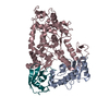
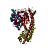
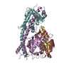
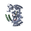
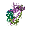
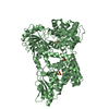
| Entry | Database: PDB / ID: 3jw0 | ||||||
|---|---|---|---|---|---|---|---|
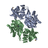
| Title | E2~Ubiquitin-HECT | ||||||
 Components Components |
| ||||||
 Keywords Keywords | LIGASE/SIGNALING PROTEIN / Ubiquitin / HECT / E3 / Ubiquitin ligase / UbcH5B / NEDD4L / NEDD4-2 / Ligase / Ubl conjugation pathway / Isopeptide bond / Nucleus / Host-virus interaction / LIGASE-SIGNALING PROTEIN complex | ||||||
| Function / homology |  Function and homology information Function and homology informationpositive regulation of caveolin-mediated endocytosis / RING-type E3 ubiquitin transferase (cysteine targeting) / negative regulation of sodium ion transmembrane transport / : / : / protein modification process => GO:0036211 / negative regulation of sodium ion import across plasma membrane / : / negative regulation of potassium ion transmembrane transport / negative regulation of potassium ion export across plasma membrane ...positive regulation of caveolin-mediated endocytosis / RING-type E3 ubiquitin transferase (cysteine targeting) / negative regulation of sodium ion transmembrane transport / : / : / protein modification process => GO:0036211 / negative regulation of sodium ion import across plasma membrane / : / negative regulation of potassium ion transmembrane transport / negative regulation of potassium ion export across plasma membrane / negative regulation of protein localization to cell surface / regulation of membrane repolarization / positive regulation of dendrite extension / receptor catabolic process / regulation of sodium ion transmembrane transport / ventricular cardiac muscle cell action potential / (E3-independent) E2 ubiquitin-conjugating enzyme / HECT-type E3 ubiquitin transferase / sodium channel inhibitor activity / potassium channel inhibitor activity / E2 ubiquitin-conjugating enzyme / regulation of dendrite morphogenesis / regulation of membrane depolarization / regulation of synapse organization / ubiquitin conjugating enzyme activity / neuromuscular junction development / sodium channel regulator activity / Peptide chain elongation / Selenocysteine synthesis / Formation of a pool of free 40S subunits / Eukaryotic Translation Termination / SRP-dependent cotranslational protein targeting to membrane / Response of EIF2AK4 (GCN2) to amino acid deficiency / Viral mRNA Translation / Nonsense Mediated Decay (NMD) independent of the Exon Junction Complex (EJC) / GTP hydrolysis and joining of the 60S ribosomal subunit / L13a-mediated translational silencing of Ceruloplasmin expression / protein autoubiquitination / Major pathway of rRNA processing in the nucleolus and cytosol / Nonsense Mediated Decay (NMD) enhanced by the Exon Junction Complex (EJC) / multivesicular body / protein K48-linked ubiquitination / Maturation of protein E / Maturation of protein E / ER Quality Control Compartment (ERQC) / Myoclonic epilepsy of Lafora / FLT3 signaling by CBL mutants / IRAK2 mediated activation of TAK1 complex / Alpha-protein kinase 1 signaling pathway / Glycogen synthesis / IRAK1 recruits IKK complex / IRAK1 recruits IKK complex upon TLR7/8 or 9 stimulation / Prevention of phagosomal-lysosomal fusion / Endosomal Sorting Complex Required For Transport (ESCRT) / Membrane binding and targetting of GAG proteins / Regulation of TBK1, IKKε (IKBKE)-mediated activation of IRF3, IRF7 / Negative regulation of FLT3 / PTK6 Regulates RTKs and Their Effectors AKT1 and DOK1 / Regulation of TBK1, IKKε-mediated activation of IRF3, IRF7 upon TLR3 ligation / IRAK2 mediated activation of TAK1 complex upon TLR7/8 or 9 stimulation / Constitutive Signaling by NOTCH1 HD Domain Mutants / NOTCH2 Activation and Transmission of Signal to the Nucleus / TICAM1,TRAF6-dependent induction of TAK1 complex / TICAM1-dependent activation of IRF3/IRF7 / APC/C:Cdc20 mediated degradation of Cyclin B / Downregulation of ERBB4 signaling / APC-Cdc20 mediated degradation of Nek2A / Regulation of FZD by ubiquitination / p75NTR recruits signalling complexes / InlA-mediated entry of Listeria monocytogenes into host cells / TRAF6 mediated IRF7 activation in TLR7/8 or 9 signaling / NF-kB is activated and signals survival / TRAF6-mediated induction of TAK1 complex within TLR4 complex / Regulation of pyruvate metabolism / Pexophagy / Downregulation of ERBB2:ERBB3 signaling / Regulation of innate immune responses to cytosolic DNA / NRIF signals cell death from the nucleus / Regulation of PTEN localization / protein modification process / VLDLR internalisation and degradation / cytosolic ribosome / Activated NOTCH1 Transmits Signal to the Nucleus / Synthesis of active ubiquitin: roles of E1 and E2 enzymes / Translesion synthesis by REV1 / TICAM1, RIP1-mediated IKK complex recruitment / Regulation of BACH1 activity / Translesion synthesis by POLK / JNK (c-Jun kinases) phosphorylation and activation mediated by activated human TAK1 / InlB-mediated entry of Listeria monocytogenes into host cell / MAP3K8 (TPL2)-dependent MAPK1/3 activation / Activation of IRF3, IRF7 mediated by TBK1, IKKε (IKBKE) / Downregulation of TGF-beta receptor signaling / Translesion synthesis by POLI / Josephin domain DUBs / Gap-filling DNA repair synthesis and ligation in GG-NER / IKK complex recruitment mediated by RIP1 / PINK1-PRKN Mediated Mitophagy / TGF-beta receptor signaling in EMT (epithelial to mesenchymal transition) / TNFR1-induced NF-kappa-B signaling pathway Similarity search - Function | ||||||
| Biological species |  Homo sapiens (human) Homo sapiens (human) | ||||||
| Method |  X-RAY DIFFRACTION / SYNCHROTRON / MOLECULAR REPLACEMENT / Resolution: 3.1 Å X-RAY DIFFRACTION / SYNCHROTRON / MOLECULAR REPLACEMENT / Resolution: 3.1 Å | ||||||
 Authors Authors | Kamadurai, H.B. / Schulman, B.A. | ||||||
 Citation Citation | Journal: Mol.Cell / Year: 2009 Title: Insights into ubiquitin transfer cascades from a structure of a UbcH5B approximately ubiquitin-HECT(NEDD4L) complex. Authors: Kamadurai, H.B. / Souphron, J. / Scott, D.C. / Duda, D.M. / Miller, D.J. / Stringer, D. / Piper, R.C. / Schulman, B.A. | ||||||
| History |
|
- Structure visualization
| Structure viewer | Molecule: MolmilJmol/JSmol |
|---|
- Downloads & links
-Download
| PDBx/mmCIF format | 3jw0.cif.gz | 241.2 KB | Display | PDBx/mmCIF format |
|---|---|---|---|---|
| PDB format | pdb3jw0.ent.gz | 194.8 KB | Display | PDB format |
| PDBx/mmJSON format | 3jw0.json.gz | Tree view | PDBx/mmJSON format | |
| Others |  Other downloads Other downloads |
-Validation report
| Arichive directory | https://data.pdbj.org/pub/pdb/validation_reports/jw/3jw0ftp://data.pdbj.org/pub/pdb/validation_reports/jw/3jw0 | HTTPS FTP |
|---|
-Related structure data


-Links
 PDBj
PDBj


- Assembly
| Deposited unit | 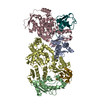
| ||||||||
|---|---|---|---|---|---|---|---|---|---|
| 1 | 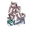
| ||||||||
| 2 | 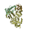
| ||||||||
| Unit cell |
| ||||||||
| Details | ACX / BDY |
-Components
| #1: Protein | Mass: 16609.963 Da / Num. of mol.: 2 / Mutation: L3S, C85S, T98K Source method: isolated from a genetically manipulated source Source: (gene. exp.) Homo sapiens (human) / Gene: UBE2D2, UBC4, UBCH5B / Production host:  #2: Protein | Mass: 45451.637 Da / Num. of mol.: 2 / Fragment: NEDD4L HECT DOMAIN (UNP 576-955) / Mutation: UBIQUITIN G76 ESTER-LINKED TO UBCH5B TO S85 Source method: isolated from a genetically manipulated source Source: (gene. exp.) Homo sapiens (human) / Gene: NEDD4L, KIAA0439, NEDL3 / Production host: References: UniProt: Q96PU5, Ligases; Forming carbon-nitrogen bonds; Acid-amino-acid ligases (peptide synthases) #3: Protein | Mass: 8922.141 Da / Num. of mol.: 2 Source method: isolated from a genetically manipulated source Source: (gene. exp.) Homo sapiens (human) / Gene: RPS27A, UBA80, UBCEP1, UBA52, UBCEP2, UBB, UBC / Production host: #4: Water | ChemComp-HOH / |  Mass: 18.015 Da / Num. of mol.: 19 / Source method: isolated from a natural source / Formula: H2O Mass: 18.015 Da / Num. of mol.: 19 / Source method: isolated from a natural source / Formula: H2OHas protein modification | Y | |
|---|
-Experimental details
-Experiment
| Experiment | Method: X-RAY DIFFRACTION / Number of used crystals: 1 |
|---|
- Sample preparation
| Crystal | Density Matthews: 3.38 Å3/Da / Density % sol: 63.59 % |
|---|---|
| Crystal grow | Temperature: 277 K / Method: vapor diffusion, hanging drop / pH: 5.1 Details: 0.1 M sodium citrate, 2.4M sodium chloride, pH 5.1, VAPOR DIFFUSION, HANGING DROP, temperature 277K |
-Data collection
| Diffraction source | Source: SYNCHROTRON / Site: APS  / Beamline: 22-ID / Wavelength: 1 Å / Beamline: 22-ID / Wavelength: 1 Å |
|---|---|
| Detector | Type: MAR scanner 300 mm plate / Detector: IMAGE PLATE / Date: Apr 20, 2008 |
| Radiation | Protocol: SINGLE WAVELENGTH / Monochromatic (M) / Laue (L): M / Scattering type: x-ray |
| Radiation wavelength | Wavelength: 1 Å / Relative weight: 1 |
| Reflection | Resolution: 3.1→50 Å / Num. obs: 34028 / Observed criterion σ(F): 0 |
- Processing
| Software |
| ||||||||||||||||||||
|---|---|---|---|---|---|---|---|---|---|---|---|---|---|---|---|---|---|---|---|---|---|
| Refinement | Method to determine structure: MOLECULAR REPLACEMENT / Resolution: 3.1→50 Å / Cross valid method: THROUGHOUT
| ||||||||||||||||||||
| Refinement step | Cycle: LAST / Resolution: 3.1→50 Å
|
