 Movie
Movie Controller

[English] 日本語
 Yorodumi
- PDB-3u3w: Crystal Structure of Bacillus thuringiensis PlcR in complex with ... -
+ Open data

- Basic information
| Entry | Database: PDB / ID: 3u3w | ||||||
|---|---|---|---|---|---|---|---|
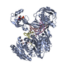
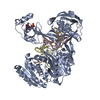
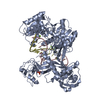
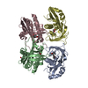
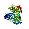
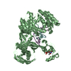
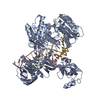
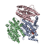
| Title | Crystal Structure of Bacillus thuringiensis PlcR in complex with the peptide PapR7 and DNA | ||||||
 Components Components |
| ||||||
 Keywords Keywords | TRANSCRIPTION ACTIVATOR/DNA / ternary complex / PlcR-PAPR7-DNA / HTH DNA-binding domain / Quorum Sensing / HTH_3 (Helix-turn-helix) domain / TPR_1 (tetratricopeptide repeats) / Pleiotropic regulator / TRANSCRIPTION ACTIVATOR-DNA complex | ||||||
| Function / homology |  Function and homology information Function and homology information | ||||||
| Biological species |  | ||||||
| Method |  X-RAY DIFFRACTION / SYNCHROTRON / MOLECULAR REPLACEMENT / Resolution: 2.4 Å X-RAY DIFFRACTION / SYNCHROTRON / MOLECULAR REPLACEMENT / Resolution: 2.4 Å | ||||||
 Authors Authors | Grenha, R. / Slamti, L. / Bouillaut, L. / Lereclus, D. / Nessler, S. | ||||||
 Citation Citation | Journal: Proc.Natl.Acad.Sci.USA / Year: 2013 Title: Structural basis for the activation mechanism of the PlcR virulence regulator by the quorum-sensing signal peptide PapR. Authors: Grenha, R. / Slamti, L. / Nicaise, M. / Refes, Y. / Lereclus, D. / Nessler, S. | ||||||
| History |
|
- Structure visualization
| Structure viewer | Molecule: MolmilJmol/JSmol |
|---|
- Downloads & links
-Download
| PDBx/mmCIF format | 3u3w.cif.gz | 162.2 KB | Display | PDBx/mmCIF format |
|---|---|---|---|---|
| PDB format | pdb3u3w.ent.gz | 123.9 KB | Display | PDB format |
| PDBx/mmJSON format | 3u3w.json.gz | Tree view | PDBx/mmJSON format | |
| Others |  Other downloads Other downloads |
-Validation report
| Arichive directory | https://data.pdbj.org/pub/pdb/validation_reports/u3/3u3wftp://data.pdbj.org/pub/pdb/validation_reports/u3/3u3w | HTTPS FTP |
|---|
-Related structure data
| Related structure data | 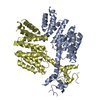 4fscC 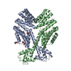 2qfcS S: Starting model for refinement C: citing same article ( |
|---|---|
| Similar structure data |

-Links
 PDBj
PDBj


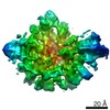
- Assembly
| Deposited unit | 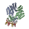
| ||||||||
|---|---|---|---|---|---|---|---|---|---|
| 1 |
| ||||||||
| Unit cell |
|
-Components
-DNA chain , 2 types, 2 molecules YZ
| #3: DNA chain | Mass: 5464.577 Da / Num. of mol.: 1 / Source method: obtained synthetically Details: PlcR box from the 1-phosphatidylinositol phosphodiesterase promoter plcA |
|---|---|
| #4: DNA chain | Mass: 5562.653 Da / Num. of mol.: 1 / Source method: obtained synthetically Details: PlcR box from the 1-phosphatidylinositol phosphodiesterase promoter plcA |
-Protein / Protein/peptide , 2 types, 4 molecules ABPQ
| #1: Protein | Mass: 35061.285 Da / Num. of mol.: 2 Source method: isolated from a genetically manipulated source Source: (gene. exp.) #2: Protein/peptide | Mass: 837.915 Da / Num. of mol.: 2 / Source method: obtained synthetically / Source: (synth.) |
|---|
-Non-polymers , 2 types, 257 molecules 

| #5: Chemical |  Mass: 40.078 Da / Num. of mol.: 2 / Source method: obtained synthetically / Formula: Ca Mass: 40.078 Da / Num. of mol.: 2 / Source method: obtained synthetically / Formula: Ca#6: Water | ChemComp-HOH / | Mass: 18.015 Da / Num. of mol.: 255 / Source method: isolated from a natural source / Formula: H2O |
|---|
-Experimental details
-Experiment
| Experiment | Method: X-RAY DIFFRACTION / Number of used crystals: 1 |
|---|
- Sample preparation
| Crystal | Density Matthews: 3.06 Å3/Da / Density % sol: 59.78 % |
|---|---|
| Crystal grow | Temperature: 291.15 K / Method: vapor diffusion, hanging drop / pH: 6.5 Details: 0.16M Calcium Acetate, 0.08M Sodium Cacodylate, 8% PEG8000, 5% Glycerol, pH 6.5, VAPOR DIFFUSION, HANGING DROP, temperature 291.15K |
-Data collection
| Diffraction | Mean temperature: 100 K | |||||||||||||||||||||||||||||||||||||||||||||||||||||||||||||||||||||||||||||
|---|---|---|---|---|---|---|---|---|---|---|---|---|---|---|---|---|---|---|---|---|---|---|---|---|---|---|---|---|---|---|---|---|---|---|---|---|---|---|---|---|---|---|---|---|---|---|---|---|---|---|---|---|---|---|---|---|---|---|---|---|---|---|---|---|---|---|---|---|---|---|---|---|---|---|---|---|---|---|
| Diffraction source | Source: SYNCHROTRON / Site: ESRF  / Beamline: ID29 / Wavelength: 0.97932 Å / Beamline: ID29 / Wavelength: 0.97932 Å | |||||||||||||||||||||||||||||||||||||||||||||||||||||||||||||||||||||||||||||
| Detector | Type: PSI PILATUS 6M / Detector: PIXEL / Date: Aug 29, 2011 / Details: cylindrical grazing incidence mirror | |||||||||||||||||||||||||||||||||||||||||||||||||||||||||||||||||||||||||||||
| Radiation | Monochromator: liquid nitrogen cooled channel-cut silicon (Si) monochromator Protocol: SINGLE WAVELENGTH / Monochromatic (M) / Laue (L): M / Scattering type: x-ray | |||||||||||||||||||||||||||||||||||||||||||||||||||||||||||||||||||||||||||||
| Radiation wavelength | Wavelength: 0.97932 Å / Relative weight: 1 | |||||||||||||||||||||||||||||||||||||||||||||||||||||||||||||||||||||||||||||
| Reflection | Resolution: 2.4→80.204 Å / Num. all: 39190 / Num. obs: 38014 / % possible obs: 97 % / Observed criterion σ(F): 0 / Observed criterion σ(I): -1 / Redundancy: 2.7 % / Biso Wilson estimate: 45.4 Å2 / Rmerge(I) obs: 0.095 / Rsym value: 0.095 / Net I/σ(I): 7.2 | |||||||||||||||||||||||||||||||||||||||||||||||||||||||||||||||||||||||||||||
| Reflection shell | Diffraction-ID: 1
|
- Processing
| Software |
| ||||||||||||||||||||||||||||||||||||||||||||||||||||||||||||||||||||||||||||||||||||||||||||||||||
|---|---|---|---|---|---|---|---|---|---|---|---|---|---|---|---|---|---|---|---|---|---|---|---|---|---|---|---|---|---|---|---|---|---|---|---|---|---|---|---|---|---|---|---|---|---|---|---|---|---|---|---|---|---|---|---|---|---|---|---|---|---|---|---|---|---|---|---|---|---|---|---|---|---|---|---|---|---|---|---|---|---|---|---|---|---|---|---|---|---|---|---|---|---|---|---|---|---|---|---|
| Refinement | Method to determine structure: MOLECULAR REPLACEMENT Starting model: PDB ENTRY 2QFC Resolution: 2.4→53.192 Å / SU ML: 0.35 / σ(F): 0 / Phase error: 23.77 / Stereochemistry target values: ML
| ||||||||||||||||||||||||||||||||||||||||||||||||||||||||||||||||||||||||||||||||||||||||||||||||||
| Solvent computation | Shrinkage radii: 0.72 Å / VDW probe radii: 1 Å / Solvent model: FLAT BULK SOLVENT MODEL / Bsol: 42.687 Å2 / ksol: 0.365 e/Å3 | ||||||||||||||||||||||||||||||||||||||||||||||||||||||||||||||||||||||||||||||||||||||||||||||||||
| Displacement parameters | Biso mean: 47.6077 Å2
| ||||||||||||||||||||||||||||||||||||||||||||||||||||||||||||||||||||||||||||||||||||||||||||||||||
| Refinement step | Cycle: LAST / Resolution: 2.4→53.192 Å
| ||||||||||||||||||||||||||||||||||||||||||||||||||||||||||||||||||||||||||||||||||||||||||||||||||
| Refine LS restraints |
| ||||||||||||||||||||||||||||||||||||||||||||||||||||||||||||||||||||||||||||||||||||||||||||||||||
| LS refinement shell | Refine-ID: X-RAY DIFFRACTION
|
