 Movie
Movie Controller

[English] 日本語
 Yorodumi
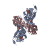
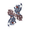
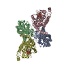
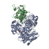
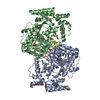
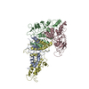
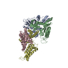
- PDB-4ii4: The Phenylacetyl-CoA monooxygenase - mutant PaaA E49Q K68Q - PaaC... -
+ Open data

- Basic information
| Entry | Database: PDB / ID: 4ii4 | ||||||
|---|---|---|---|---|---|---|---|
| Title | The Phenylacetyl-CoA monooxygenase - mutant PaaA E49Q K68Q - PaaC wild type subcomplex with benzoyl-CoA | ||||||
 Components Components |
| ||||||
 Keywords Keywords | OXIDOREDUCTASE / protein-protein complex / Structural Genomics / Montreal-Kingston Bacterial Structural Genomics Initiative / BSGI / ferritin-like fold / bacterial multicomponent monooxygenase PaaABCE | ||||||
| Function / homology |  Function and homology information Function and homology informationphenylacetyl-CoA 1,2-epoxidase / phenylacetyl-CoA 1,2-epoxidase complex / phenylacetyl-CoA 1,2-epoxidase activity / phenylacetate catabolic process / cytosol Similarity search - Function | ||||||
| Biological species |  | ||||||
| Method |  X-RAY DIFFRACTION / SYNCHROTRON / isomorphous replacement with 3PW1 / Resolution: 2.799 Å X-RAY DIFFRACTION / SYNCHROTRON / isomorphous replacement with 3PW1 / Resolution: 2.799 Å | ||||||
 Authors Authors | Cygler, M. / Grishin, A.M. / Montreal-Kingston Bacterial Structural Genomics Initiative (BSGI) | ||||||
 Citation Citation | Journal: To be published Title: Spatial Organization of Subunits within the Phenylacetyl-CoA Monooxygenase Complex Authors: Grishin, A.M. / Ajamian, E. / Tao, L. / Bostina, M. / Zhang, L. / Trempe, J. / Menard, R. / Rouiller, I. / Cygler, M. | ||||||
| History |
|
- Structure visualization
| Structure viewer | Molecule: MolmilJmol/JSmol |
|---|
- Downloads & links
-Download
| PDBx/mmCIF format | 4ii4.cif.gz | 335.2 KB | Display | PDBx/mmCIF format |
|---|---|---|---|---|
| PDB format | pdb4ii4.ent.gz | 274.8 KB | Display | PDB format |
| PDBx/mmJSON format | 4ii4.json.gz | Tree view | PDBx/mmJSON format | |
| Others |  Other downloads Other downloads |
-Validation report
| Arichive directory | https://data.pdbj.org/pub/pdb/validation_reports/ii/4ii4ftp://data.pdbj.org/pub/pdb/validation_reports/ii/4ii4 | HTTPS FTP |
|---|
-Related structure data
| Related structure data | 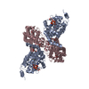 3pw1S S: Starting model for refinement |
|---|---|
| Similar structure data | |
| Other databases |

-Links
 PDBj
PDBj


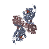
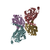
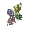
- Assembly
| Deposited unit | 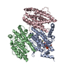
| ||||||||
|---|---|---|---|---|---|---|---|---|---|
| 1 | 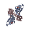
| ||||||||
| 2 | 
| ||||||||
| Unit cell |
|
-Components
| #1: Protein | Mass: 35786.500 Da / Num. of mol.: 1 / Mutation: E49Q, K68Q Source method: isolated from a genetically manipulated source Details: paaA gene contains mutations that lead to aminoacid changes E49Q, K68Q Source: (gene. exp.) | ||||
|---|---|---|---|---|---|
| #2: Protein | Mass: 29109.629 Da / Num. of mol.: 2 Source method: isolated from a genetically manipulated source Source: (gene. exp.) References: UniProt: P76079, Oxidoreductases; Acting on paired donors, with incorporation or reduction of molecular oxygen; With NADH or NADPH as one donor, and incorporation of one atom of oxygen into the other donor #3: Chemical | ChemComp-BYC / | 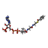  Mass: 871.640 Da / Num. of mol.: 1 / Source method: obtained synthetically / Formula: C28H40N7O17P3S Mass: 871.640 Da / Num. of mol.: 1 / Source method: obtained synthetically / Formula: C28H40N7O17P3S#4: Water | ChemComp-HOH / |  Mass: 18.015 Da / Num. of mol.: 25 / Source method: isolated from a natural source / Formula: H2O Mass: 18.015 Da / Num. of mol.: 25 / Source method: isolated from a natural source / Formula: H2O |
-Experimental details
-Experiment
| Experiment | Method: X-RAY DIFFRACTION / Number of used crystals: 1 |
|---|
- Sample preparation
| Crystal | Density Matthews: 2.44 Å3/Da / Density % sol: 49.58 % |
|---|---|
| Crystal grow | Temperature: 293 K / Method: vapor diffusion, hanging drop / pH: 8 Details: 25% ethylene glycol, pH 8.0, VAPOR DIFFUSION, HANGING DROP, temperature 293K |
-Data collection
| Diffraction | Mean temperature: 77.2 K | |||||||||||||||||||||||||||||||||||||||||||||||||||||||||||||||||||||||||||||||||||||||||||||||||||||||||||||||||||||||||||||||||||||||||||||||||||
|---|---|---|---|---|---|---|---|---|---|---|---|---|---|---|---|---|---|---|---|---|---|---|---|---|---|---|---|---|---|---|---|---|---|---|---|---|---|---|---|---|---|---|---|---|---|---|---|---|---|---|---|---|---|---|---|---|---|---|---|---|---|---|---|---|---|---|---|---|---|---|---|---|---|---|---|---|---|---|---|---|---|---|---|---|---|---|---|---|---|---|---|---|---|---|---|---|---|---|---|---|---|---|---|---|---|---|---|---|---|---|---|---|---|---|---|---|---|---|---|---|---|---|---|---|---|---|---|---|---|---|---|---|---|---|---|---|---|---|---|---|---|---|---|---|---|---|---|---|
| Diffraction source | Source: SYNCHROTRON / Site: CLSI  / Beamline: 08ID-1 / Wavelength: 0.97949 Å / Beamline: 08ID-1 / Wavelength: 0.97949 Å | |||||||||||||||||||||||||||||||||||||||||||||||||||||||||||||||||||||||||||||||||||||||||||||||||||||||||||||||||||||||||||||||||||||||||||||||||||
| Detector | Type: RAYONIX MX-300 / Detector: CCD / Date: Dec 10, 2010 | |||||||||||||||||||||||||||||||||||||||||||||||||||||||||||||||||||||||||||||||||||||||||||||||||||||||||||||||||||||||||||||||||||||||||||||||||||
| Radiation | Monochromator: dcm WITH CRYO-COOLED 1ST CRYSTAL SAGITALLY BENT 2ND CRYSTAL FOLLOWED BY VERTICALLY FOCUSING MIRROR Protocol: SINGLE WAVELENGTH / Monochromatic (M) / Laue (L): M / Scattering type: x-ray | |||||||||||||||||||||||||||||||||||||||||||||||||||||||||||||||||||||||||||||||||||||||||||||||||||||||||||||||||||||||||||||||||||||||||||||||||||
| Radiation wavelength | Wavelength: 0.97949 Å / Relative weight: 1 | |||||||||||||||||||||||||||||||||||||||||||||||||||||||||||||||||||||||||||||||||||||||||||||||||||||||||||||||||||||||||||||||||||||||||||||||||||
| Reflection | Resolution: 2.8→50 Å / Num. obs: 24054 / % possible obs: 99.8 % / Redundancy: 14 % / Rmerge(I) obs: 0.127 / Χ2: 1.709 / Net I/σ(I): 5.6 | |||||||||||||||||||||||||||||||||||||||||||||||||||||||||||||||||||||||||||||||||||||||||||||||||||||||||||||||||||||||||||||||||||||||||||||||||||
| Reflection shell |
|
- Processing
| Software |
| |||||||||||||||||||||||||||||||||||||||||||||||||||||||||||||||||||||||||||||||||||||||||||||||||||||||||||||||||||||||||||||
|---|---|---|---|---|---|---|---|---|---|---|---|---|---|---|---|---|---|---|---|---|---|---|---|---|---|---|---|---|---|---|---|---|---|---|---|---|---|---|---|---|---|---|---|---|---|---|---|---|---|---|---|---|---|---|---|---|---|---|---|---|---|---|---|---|---|---|---|---|---|---|---|---|---|---|---|---|---|---|---|---|---|---|---|---|---|---|---|---|---|---|---|---|---|---|---|---|---|---|---|---|---|---|---|---|---|---|---|---|---|---|---|---|---|---|---|---|---|---|---|---|---|---|---|---|---|---|
| Refinement | Method to determine structure: isomorphous replacement with 3PW1 Starting model: 3PW1 Resolution: 2.799→26.638 Å / Occupancy max: 1 / Occupancy min: 1 / SU ML: 0.48 / σ(F): 1.33 / Phase error: 32.32 / Stereochemistry target values: ML
| |||||||||||||||||||||||||||||||||||||||||||||||||||||||||||||||||||||||||||||||||||||||||||||||||||||||||||||||||||||||||||||
| Solvent computation | Shrinkage radii: 0.9 Å / VDW probe radii: 1.11 Å / Solvent model: FLAT BULK SOLVENT MODEL | |||||||||||||||||||||||||||||||||||||||||||||||||||||||||||||||||||||||||||||||||||||||||||||||||||||||||||||||||||||||||||||
| Displacement parameters | Biso max: 221.41 Å2 / Biso mean: 119.142 Å2 / Biso min: 46.84 Å2 | |||||||||||||||||||||||||||||||||||||||||||||||||||||||||||||||||||||||||||||||||||||||||||||||||||||||||||||||||||||||||||||
| Refinement step | Cycle: LAST / Resolution: 2.799→26.638 Å
| |||||||||||||||||||||||||||||||||||||||||||||||||||||||||||||||||||||||||||||||||||||||||||||||||||||||||||||||||||||||||||||
| Refinement TLS params. | Method: refined / Refine-ID: X-RAY DIFFRACTION
| |||||||||||||||||||||||||||||||||||||||||||||||||||||||||||||||||||||||||||||||||||||||||||||||||||||||||||||||||||||||||||||
| Refinement TLS group |
|
