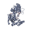
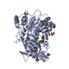
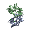
- PDB-4hcc: The zinc ion bound form of crystal structure of E.coli ExoI-ssDNA... -
+
Open data
ID or keywords:
Loading...
-
Basic information
Entry
Database: PDB / ID: 4hcc
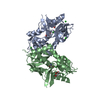
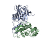
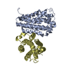
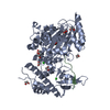
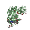
Title
The zinc ion bound form of crystal structure of E.coli ExoI-ssDNA complex
Components
DNA (5'-D(*AP*AP*AP*AP*AP*AP*AP*AP*AP*AP*AP*A)-3')
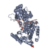
Exodeoxyribonuclease I
Keywords
HYDROLASE/DNA / DnaQ family / Exonuclease C-terminal family / HYDROLASE-DNA complex
Function / homology
Function and homology information
exodeoxyribonuclease I / DNA replication termination / single-stranded DNA 3'-5' DNA exonuclease activity / DNA catabolic process / 5'-deoxyribose-5-phosphate lyase activity / single-stranded DNA binding / 3'-5'-RNA exonuclease activity / DNA repair / magnesium ion binding Similarity search - Function
: / Exonuclease I C-terminal domain / Helix Hairpins - #1240 / Exonuclease ExoI, domain 3 / Exonuclease ExoI, domain 2 / Exodeoxyribonuclease I, C-terminal / Exodeoxyribonuclease I / Exonuclease I, SH3-like domain / Exonuclease I, SH3-like domain superfamily / Exonuclease I SH3-like domain ...: / Exonuclease I C-terminal domain / Helix Hairpins - #1240 / Exonuclease ExoI, domain 3 / Exonuclease ExoI, domain 2 / Exodeoxyribonuclease I, C-terminal / Exodeoxyribonuclease I / Exonuclease I, SH3-like domain / Exonuclease I, SH3-like domain superfamily / Exonuclease I SH3-like domain / Exonuclease I (ExoI) SH3-like domain profile. / Exonuclease I (ExoI) C-terminal domain profile. / Oligoribonuclease / PX Domain / Exonuclease / Exonuclease, RNase T/DNA polymerase III / EXOIII / Monooxygenase / Ribonuclease H-like superfamily/Ribonuclease H / Nucleotidyltransferase; domain 5 / Helix Hairpins / Ribonuclease H superfamily / Ribonuclease H-like superfamily / Up-down Bundle / 2-Layer Sandwich / Orthogonal Bundle / Mainly Alpha / Alpha Beta Similarity search - Domain/homology
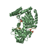
A: Exodeoxyribonuclease I B: Exodeoxyribonuclease I C: DNA (5'-D(*AP*AP*AP*AP*AP*AP*AP*AP*AP*AP*AP*A)-3') D: DNA (5'-D(*AP*AP*AP*AP*AP*AP*AP*AP*AP*AP*AP*A)-3') hetero molecules
ExodeoxyribonucleaseI / Exonuclease I / DNA deoxyribophosphodiesterase / dRPase
Mass: 55381.453 Da / Num. of mol.: 2 Source method: isolated from a genetically manipulated source Source: (gene. exp.) Escherichia coli (E. coli) / Strain: DH5alpha / Gene: b2011, cpeA, JW1993, sbcB, xonA / Plasmid: pET22b / Production host: Escherichia coli (E. coli) / Strain (production host): BL21(DE3) / References: UniProt: P04995, exodeoxyribonuclease I
#2: DNA chain
DNA (5'-D(*AP*AP*AP*AP*AP*AP*AP*AP*AP*AP*AP*A)-3')
Mass: 3713.524 Da / Num. of mol.: 2 / Source method: obtained synthetically Details: The ssDNA samples were synthesized with 3'-phosphoryl modification (PAGE purified)
Mass: 18.015 Da / Num. of mol.: 20 / Source method: isolated from a natural source / Formula: H2O
-
Details
Sequence details
THIS RESIDUE IS ASP FOR E.COLI STRAIN DH5ALPHA
-
Experimental details
-
Experiment
Experiment
Method: X-RAY DIFFRACTION / Number of used crystals: 1
-
Sample preparation
Crystal
Density Matthews: 2.3 Å3/Da / Density % sol: 46.42 %
Crystal grow
Temperature: 293 K / Method: vapor diffusion, sitting drop / pH: 6 Details: The crystals grew in 1.65-1.68 M ammonium sulfate, 6.5-6.8% v/v 2-propanol, 0.1 M sodium citrate tribasic dihydrate pH 6.0, 5 mM phenol, then soaked in the solution consisted of precipitant ...Details: The crystals grew in 1.65-1.68 M ammonium sulfate, 6.5-6.8% v/v 2-propanol, 0.1 M sodium citrate tribasic dihydrate pH 6.0, 5 mM phenol, then soaked in the solution consisted of precipitant solution and 20 mM zinc chloride, VAPOR DIFFUSION, SITTING DROP, temperature 293K
Resolution: 2.96→30 Å / Cor.coef. Fo:Fc: 0.941 / Cor.coef. Fo:Fc free: 0.896 / Occupancy max: 1 / Occupancy min: 0 / SU B: 16.616 / SU ML: 0.31 / Cross valid method: THROUGHOUT / σ(F): 0 / ESU R Free: 0.448 / Stereochemistry target values: MAXIMUM LIKELIHOOD Details: HYDROGENS HAVE BEEN USED IF PRESENT IN THE INPUT U VALUES: REFINED INDIVIDUALLY
Rfactor
Num. reflection
% reflection
Selection details
Rfree
0.2448
1188
5.1 %
RANDOM
Rwork
0.1849
-
-
-
obs
0.188
23099
98.49 %
-
all
-
23347
-
-
Solvent computation
Ion probe radii: 0.8 Å / Shrinkage radii: 0.8 Å / VDW probe radii: 1.2 Å / Solvent model: MASK
In the structure databanks used in Yorodumi, some data are registered as the other names, "COVID-19 virus" and "2019-nCoV". Here are the details of the virus and the list of structure data.
Jan 31, 2019. EMDB accession codes are about to change! (news from PDBe EMDB page)
EMDB accession codes are about to change! (news from PDBe EMDB page)
The allocation of 4 digits for EMDB accession codes will soon come to an end. Whilst these codes will remain in use, new EMDB accession codes will include an additional digit and will expand incrementally as the available range of codes is exhausted. The current 4-digit format prefixed with “EMD-” (i.e. EMD-XXXX) will advance to a 5-digit format (i.e. EMD-XXXXX), and so on. It is currently estimated that the 4-digit codes will be depleted around Spring 2019, at which point the 5-digit format will come into force.
The EM Navigator/Yorodumi systems omit the EMD- prefix.
Related info.:Q: What is EMD? / ID/Accession-code notation in Yorodumi/EM Navigator
Yorodumi is a browser for structure data from EMDB, PDB, SASBDB, etc.
This page is also the successor to EM Navigator detail page, and also detail information page/front-end page for Omokage search.
The word "yorodu" (or yorozu) is an old Japanese word meaning "ten thousand". "mi" (miru) is to see.
Related info.:EMDB / PDB / SASBDB / Comparison of 3 databanks / Yorodumi Search / Aug 31, 2016. New EM Navigator & Yorodumi / Yorodumi Papers / Jmol/JSmol / Function and homology information / Changes in new EM Navigator and Yorodumi
 Movie
Movie Controller

 Yorodumi
 Open data
 Basic information
 Components
 Keywords
 Function and homology information
 X-RAY DIFFRACTION /
  Authors
 Citation
 Structure visualization
 Downloads & links
 Other downloads
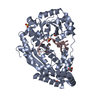


 PDBj
PDBj


 Assembly
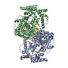


 Mass: 96.063 Da / Num. of mol.: 7 / Source method: obtained synthetically / Formula: SO4
 Mass: 65.409 Da / Num. of mol.: 2 / Source method: obtained synthetically / Formula: Zn
 Mass: 60.095 Da / Num. of mol.: 3 / Source method: obtained synthetically / Formula: C3H8O
 Mass: 94.971 Da / Num. of mol.: 2 / Source method: obtained synthetically / Formula: PO4
 Sample preparation
 / Beamline: BL17U / Wavelength: 0.9793 Å
 Processing