 Movie
Movie Controller

[English] 日本語
 Yorodumi
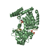
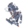
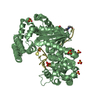
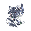
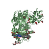
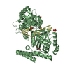
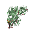
- PDB-4js5: Crystal structure of E. coli Exonuclease I in complex with a dT13... -
+ Open data

- Basic information
| Entry | Database: PDB / ID: 4js5 | ||||||
|---|---|---|---|---|---|---|---|
| Title | Crystal structure of E. coli Exonuclease I in complex with a dT13 oligonucleotide | ||||||
 Components Components |
| ||||||
 Keywords Keywords | HYDROLASE/DNA / DNA repair / exonuclease / processive / DnaQ superfamily / 3'-5' ssDNA exonuclease / HYDROLASE / HYDROLASE-DNA complex | ||||||
| Function / homology |  Function and homology information Function and homology informationexodeoxyribonuclease I / DNA replication termination / single-stranded DNA 3'-5' DNA exonuclease activity / DNA catabolic process / 5'-deoxyribose-5-phosphate lyase activity / single-stranded DNA binding / 3'-5'-RNA exonuclease activity / DNA repair / magnesium ion binding Similarity search - Function | ||||||
| Biological species |  | ||||||
| Method |  X-RAY DIFFRACTION / SYNCHROTRON / MOLECULAR REPLACEMENT / Resolution: 3.5 Å X-RAY DIFFRACTION / SYNCHROTRON / MOLECULAR REPLACEMENT / Resolution: 3.5 Å | ||||||
 Authors Authors | Bell, C.E. | ||||||
 Citation Citation | Journal: Nucleic Acids Res. / Year: 2013 Title: Crystal structures of Escherichia coli exonuclease I in complex with single-stranded DNA provide insights into the mechanism of processive digestion. Authors: Korada, S.K. / Johns, T.D. / Smith, C.E. / Jones, N.D. / McCabe, K.A. / Bell, C.E. | ||||||
| History |
|
- Structure visualization
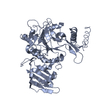
| Structure viewer | Molecule: MolmilJmol/JSmol |
|---|
- Downloads & links
-Download
| PDBx/mmCIF format | 4js5.cif.gz | 205.4 KB | Display | PDBx/mmCIF format |
|---|---|---|---|---|
| PDB format | pdb4js5.ent.gz | 164.9 KB | Display | PDB format |
| PDBx/mmJSON format | 4js5.json.gz | Tree view | PDBx/mmJSON format | |
| Others |  Other downloads Other downloads |
-Validation report
| Arichive directory | https://data.pdbj.org/pub/pdb/validation_reports/js/4js5ftp://data.pdbj.org/pub/pdb/validation_reports/js/4js5 | HTTPS FTP |
|---|
-Related structure data
| Related structure data | 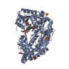 4jrpC 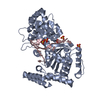 4jrqC 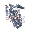 4js4C 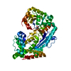 1fxxS C: citing same article ( S: Starting model for refinement |
|---|---|
| Similar structure data |


-Links
 PDBj
PDBj


- Assembly
| Deposited unit | 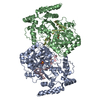
| ||||||||||||
|---|---|---|---|---|---|---|---|---|---|---|---|---|---|
| 1 | 
| ||||||||||||
| 2 | 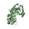
| ||||||||||||
| Unit cell |
| ||||||||||||
| Noncrystallographic symmetry (NCS) | NCS oper:
|
-Components
| #1: DNA chain | Mass: 3909.549 Da / Num. of mol.: 2 / Source method: obtained synthetically / Details: chemical synthesis #2: Protein | Mass: 54848.891 Da / Num. of mol.: 2 Source method: isolated from a genetically manipulated source Details: N-terminal 6His-thrombin tab / Source: (gene. exp.) #3: Chemical |   Mass: 96.063 Da / Num. of mol.: 2 / Source method: obtained synthetically / Formula: SO4 Mass: 96.063 Da / Num. of mol.: 2 / Source method: obtained synthetically / Formula: SO4 |
|---|
-Experimental details
-Experiment
| Experiment | Method: X-RAY DIFFRACTION / Number of used crystals: 1 |
|---|
- Sample preparation
| Crystal | Density Matthews: 4.65 Å3/Da / Density % sol: 73.55 % |
|---|---|
| Crystal grow | Temperature: 298 K / Method: vapor diffusion, hanging drop / pH: 8 Details: 5% 2-propanol, 25 % glycerol, 1.2 M ammonium sulfate, pH 8, VAPOR DIFFUSION, HANGING DROP, temperature 298K |
-Data collection
| Diffraction | Mean temperature: 100 K |
|---|---|
| Diffraction source | Source: SYNCHROTRON / Site: APS  / Beamline: 31-ID / Wavelength: 0.97931 Å / Beamline: 31-ID / Wavelength: 0.97931 Å |
| Detector | Type: RAYONIX MX225HE / Detector: CCD / Date: Nov 16, 2012 |
| Radiation | Monochromator: Kohzu HLD-4 double crystal / Protocol: SINGLE WAVELENGTH / Monochromatic (M) / Laue (L): M / Scattering type: x-ray |
| Radiation wavelength | Wavelength: 0.97931 Å / Relative weight: 1 |
| Reflection | Resolution: 3.5→49 Å / Num. all: 26653 / Num. obs: 26653 / % possible obs: 99.9 % / Observed criterion σ(F): 1 / Observed criterion σ(I): 1 / Redundancy: 11 % / Rmerge(I) obs: 0.1 / Net I/σ(I): 9.6 |
| Reflection shell | Resolution: 3.5→3.69 Å / Redundancy: 10.1 % / Rmerge(I) obs: 0.786 / Mean I/σ(I) obs: 2 / % possible all: 100 |
- Processing
| Software |
| ||||||||||||||||||||||||||||||||||||||||||||||||||||||||||||||||||||||||||||||||||||||||||||||||||||||||||||||||||||||||||||||||||||||||||||||||||||||||||||||||||||||||||
|---|---|---|---|---|---|---|---|---|---|---|---|---|---|---|---|---|---|---|---|---|---|---|---|---|---|---|---|---|---|---|---|---|---|---|---|---|---|---|---|---|---|---|---|---|---|---|---|---|---|---|---|---|---|---|---|---|---|---|---|---|---|---|---|---|---|---|---|---|---|---|---|---|---|---|---|---|---|---|---|---|---|---|---|---|---|---|---|---|---|---|---|---|---|---|---|---|---|---|---|---|---|---|---|---|---|---|---|---|---|---|---|---|---|---|---|---|---|---|---|---|---|---|---|---|---|---|---|---|---|---|---|---|---|---|---|---|---|---|---|---|---|---|---|---|---|---|---|---|---|---|---|---|---|---|---|---|---|---|---|---|---|---|---|---|---|---|---|---|---|---|---|
| Refinement | Method to determine structure: MOLECULAR REPLACEMENT Starting model: PDB ENTRY 1FXX Resolution: 3.5→49 Å / Cor.coef. Fo:Fc: 0.93 / Cor.coef. Fo:Fc free: 0.909 / SU B: 61.309 / SU ML: 0.789 / Cross valid method: THROUGHOUT / σ(F): 1 / σ(I): 1 / ESU R Free: 0.67 / Stereochemistry target values: MAXIMUM LIKELIHOOD / Details: HYDROGENS HAVE BEEN ADDED IN THE RIDING POSITIONS
| ||||||||||||||||||||||||||||||||||||||||||||||||||||||||||||||||||||||||||||||||||||||||||||||||||||||||||||||||||||||||||||||||||||||||||||||||||||||||||||||||||||||||||
| Solvent computation | Ion probe radii: 0.8 Å / Shrinkage radii: 0.8 Å / VDW probe radii: 1.2 Å / Solvent model: MASK | ||||||||||||||||||||||||||||||||||||||||||||||||||||||||||||||||||||||||||||||||||||||||||||||||||||||||||||||||||||||||||||||||||||||||||||||||||||||||||||||||||||||||||
| Displacement parameters | Biso mean: 196.357 Å2
| ||||||||||||||||||||||||||||||||||||||||||||||||||||||||||||||||||||||||||||||||||||||||||||||||||||||||||||||||||||||||||||||||||||||||||||||||||||||||||||||||||||||||||
| Refinement step | Cycle: LAST / Resolution: 3.5→49 Å
| ||||||||||||||||||||||||||||||||||||||||||||||||||||||||||||||||||||||||||||||||||||||||||||||||||||||||||||||||||||||||||||||||||||||||||||||||||||||||||||||||||||||||||
| Refine LS restraints |
| ||||||||||||||||||||||||||||||||||||||||||||||||||||||||||||||||||||||||||||||||||||||||||||||||||||||||||||||||||||||||||||||||||||||||||||||||||||||||||||||||||||||||||
| LS refinement shell | Resolution: 3.5→3.591 Å / Total num. of bins used: 20
|
