 Movie
Movie Controller

[English] 日本語
 Yorodumi
- PDB-4io4: Crystal Structure of the AvGluR1 ligand binding domain complex wi... -
+ Open data

- Basic information
| Entry | Database: PDB / ID: 4io4 | ||||||
|---|---|---|---|---|---|---|---|
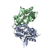
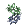
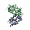
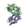
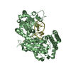
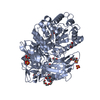
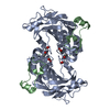
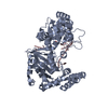
| Title | Crystal Structure of the AvGluR1 ligand binding domain complex with serine at 1.94 Angstrom resolution | ||||||
 Components Components | AvGluR1 ligand binding domain | ||||||
 Keywords Keywords | MEMBRANE PROTEIN | ||||||
| Function / homology |  Function and homology information Function and homology informationligand-gated monoatomic ion channel activity / G protein-coupled receptor activity / postsynaptic membrane Similarity search - Function | ||||||
| Biological species |  Adineta vaga (invertebrata) Adineta vaga (invertebrata) | ||||||
| Method |  X-RAY DIFFRACTION / SYNCHROTRON / MOLECULAR REPLACEMENT / Resolution: 1.941 Å X-RAY DIFFRACTION / SYNCHROTRON / MOLECULAR REPLACEMENT / Resolution: 1.941 Å | ||||||
 Authors Authors | Lomash, S. / Chittori, S. / Mayer, M.L. | ||||||
 Citation Citation | Journal: Structure / Year: 2013 Title: Anions Mediate Ligand Binding in Adineta vaga Glutamate Receptor Ion Channels. Authors: Lomash, S. / Chittori, S. / Brown, P. / Mayer, M.L. | ||||||
| History |
|
- Structure visualization
| Structure viewer | Molecule: MolmilJmol/JSmol |
|---|
- Downloads & links
-Download
| PDBx/mmCIF format | 4io4.cif.gz | 302 KB | Display | PDBx/mmCIF format |
|---|---|---|---|---|
| PDB format | pdb4io4.ent.gz | 252.5 KB | Display | PDB format |
| PDBx/mmJSON format | 4io4.json.gz | Tree view | PDBx/mmJSON format | |
| Others |  Other downloads Other downloads |
-Validation report
| Arichive directory | https://data.pdbj.org/pub/pdb/validation_reports/io/4io4ftp://data.pdbj.org/pub/pdb/validation_reports/io/4io4 | HTTPS FTP |
|---|
-Related structure data
| Related structure data |  4io2SC  4io3C  4io5C  4io6C  4io7C S: Starting model for refinement C: citing same article ( |
|---|---|
| Similar structure data |

-Links
 PDBj
PDBj

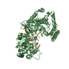
- Assembly
| Deposited unit | 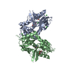
| ||||||||
|---|---|---|---|---|---|---|---|---|---|
| 1 |
| ||||||||
| Unit cell |
|
-Components
| #1: Protein | Mass: 27492.455 Da / Num. of mol.: 2 / Fragment: unp residues 457-567; 680-812 Source method: isolated from a genetically manipulated source Source: (gene. exp.) Adineta vaga (invertebrata) / Plasmid: pet22B modified / Production host:  #2: Chemical |   Type: L-peptide linking / Mass: 105.093 Da / Num. of mol.: 2 / Source method: obtained synthetically / Formula: C3H7NO3 Type: L-peptide linking / Mass: 105.093 Da / Num. of mol.: 2 / Source method: obtained synthetically / Formula: C3H7NO3#3: Chemical | ChemComp-CL /   Mass: 35.453 Da / Num. of mol.: 7 / Source method: obtained synthetically / Formula: Cl Mass: 35.453 Da / Num. of mol.: 7 / Source method: obtained synthetically / Formula: Cl#4: Chemical |   Mass: 92.094 Da / Num. of mol.: 2 / Source method: obtained synthetically / Formula: C3H8O3 Mass: 92.094 Da / Num. of mol.: 2 / Source method: obtained synthetically / Formula: C3H8O3#5: Water | ChemComp-HOH / |  Mass: 18.015 Da / Num. of mol.: 262 / Source method: isolated from a natural source / Formula: H2O Mass: 18.015 Da / Num. of mol.: 262 / Source method: isolated from a natural source / Formula: H2OHas protein modification | Y | Sequence details | THE PROTEIN CRYSTALLIZED IS THE EXTRACELLULAR LIGAND BINDING DOMAIN OF AvGluR1. TRANSMEMBRANE ...THE PROTEIN CRYSTALLIZ | |
|---|
-Experimental details
-Experiment
| Experiment | Method: X-RAY DIFFRACTION / Number of used crystals: 1 |
|---|
- Sample preparation
| Crystal | Density Matthews: 2.68 Å3/Da / Density % sol: 54.19 % |
|---|---|
| Crystal grow | Temperature: 293 K / Method: vapor diffusion, hanging drop / pH: 6.5 Details: 20% PEG 3350, 0.1 M NaCitrate, 0.1 M BisTris Propane, pH 6.5, VAPOR DIFFUSION, HANGING DROP, temperature 293K |
-Data collection
| Diffraction source | Source: SYNCHROTRON / Site: APS  / Beamline: 22-ID / Wavelength: 1 Å / Beamline: 22-ID / Wavelength: 1 Å |
|---|---|
| Detector | Type: MARMOSAIC 300 mm CCD / Detector: CCD / Date: Apr 23, 2012 |
| Radiation | Monochromator: SI(111) / Protocol: SINGLE WAVELENGTH / Monochromatic (M) / Laue (L): M / Scattering type: x-ray |
| Radiation wavelength | Wavelength: 1 Å / Relative weight: 1 |
| Reflection | Resolution: 1.94→40 Å / Num. all: 43189 / Num. obs: 43189 / % possible obs: 99.9 % / Observed criterion σ(F): 0 / Observed criterion σ(I): 0 / Redundancy: 3.8 % / Biso Wilson estimate: 29.5 Å2 / Rmerge(I) obs: 0.05 / Net I/σ(I): 27.4 |
| Reflection shell | Resolution: 1.94→1.97 Å / Redundancy: 3.8 % / Rmerge(I) obs: 0.56 / Mean I/σ(I) obs: 2.4 / % possible all: 100 |
- Processing
| Software |
| |||||||||||||||||||||||||||||||||||||||||||||||||||||||||||||||||||||||||||||||||||||||||||||||||||||||||||||||||||||||||||||||||||||||||||||||||||||||||||||||||||||||||||||||||||||||||||||||||||||||||||||||||||||||||||||||||
|---|---|---|---|---|---|---|---|---|---|---|---|---|---|---|---|---|---|---|---|---|---|---|---|---|---|---|---|---|---|---|---|---|---|---|---|---|---|---|---|---|---|---|---|---|---|---|---|---|---|---|---|---|---|---|---|---|---|---|---|---|---|---|---|---|---|---|---|---|---|---|---|---|---|---|---|---|---|---|---|---|---|---|---|---|---|---|---|---|---|---|---|---|---|---|---|---|---|---|---|---|---|---|---|---|---|---|---|---|---|---|---|---|---|---|---|---|---|---|---|---|---|---|---|---|---|---|---|---|---|---|---|---|---|---|---|---|---|---|---|---|---|---|---|---|---|---|---|---|---|---|---|---|---|---|---|---|---|---|---|---|---|---|---|---|---|---|---|---|---|---|---|---|---|---|---|---|---|---|---|---|---|---|---|---|---|---|---|---|---|---|---|---|---|---|---|---|---|---|---|---|---|---|---|---|---|---|---|---|---|---|---|---|---|---|---|---|---|---|---|---|---|---|---|---|---|---|
| Refinement | Method to determine structure: MOLECULAR REPLACEMENT Starting model: 4Io2 Resolution: 1.941→35.062 Å / SU ML: 0.15 / Cross valid method: THROUGHOUT / σ(F): 1.36 / Phase error: 19.41 / Stereochemistry target values: ML
| |||||||||||||||||||||||||||||||||||||||||||||||||||||||||||||||||||||||||||||||||||||||||||||||||||||||||||||||||||||||||||||||||||||||||||||||||||||||||||||||||||||||||||||||||||||||||||||||||||||||||||||||||||||||||||||||||
| Solvent computation | Shrinkage radii: 0.9 Å / VDW probe radii: 1.11 Å / Solvent model: FLAT BULK SOLVENT MODEL | |||||||||||||||||||||||||||||||||||||||||||||||||||||||||||||||||||||||||||||||||||||||||||||||||||||||||||||||||||||||||||||||||||||||||||||||||||||||||||||||||||||||||||||||||||||||||||||||||||||||||||||||||||||||||||||||||
| Refinement step | Cycle: LAST / Resolution: 1.941→35.062 Å
| |||||||||||||||||||||||||||||||||||||||||||||||||||||||||||||||||||||||||||||||||||||||||||||||||||||||||||||||||||||||||||||||||||||||||||||||||||||||||||||||||||||||||||||||||||||||||||||||||||||||||||||||||||||||||||||||||
| Refine LS restraints |
| |||||||||||||||||||||||||||||||||||||||||||||||||||||||||||||||||||||||||||||||||||||||||||||||||||||||||||||||||||||||||||||||||||||||||||||||||||||||||||||||||||||||||||||||||||||||||||||||||||||||||||||||||||||||||||||||||
| LS refinement shell |
| |||||||||||||||||||||||||||||||||||||||||||||||||||||||||||||||||||||||||||||||||||||||||||||||||||||||||||||||||||||||||||||||||||||||||||||||||||||||||||||||||||||||||||||||||||||||||||||||||||||||||||||||||||||||||||||||||
| Refinement TLS params. | Method: refined / Refine-ID: X-RAY DIFFRACTION
| |||||||||||||||||||||||||||||||||||||||||||||||||||||||||||||||||||||||||||||||||||||||||||||||||||||||||||||||||||||||||||||||||||||||||||||||||||||||||||||||||||||||||||||||||||||||||||||||||||||||||||||||||||||||||||||||||
| Refinement TLS group |
|
