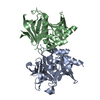
Entry Database : PDB / ID : 1mk2Title SMAD3 SBD complex Madh-interacting protein SMAD 3 Keywords / / / Function / homology Function Domain/homology Component
/ / / / / / / / / / / / / / / / / / / / / / / / / / / / / / / / / / / / / / / / / / / / / / / / / / / / / / / / / / / / / / / / / / / / / / / / / / / / / / / / / / / / / / / / / / / / / / / / / / / / / / / / / / / / / / / / / / / / / / / / / / / / / / / / / / / / / / / / / / / / / / / / / / / / Biological species Homo sapiens (human)Method / / / Resolution : 2.74 Å Authors Qin, B.Y. / Lam, S.S. / Correia, J.J. / Lin, K. Journal : Genes Dev. / Year : 2002Title : Smad3 allostery links TGF-beta receptor kinase activation to transcriptional controlAuthors : Qin, B.Y. / Lam, S.S. / Correia, J.J. / Lin, K. History Deposition Aug 28, 2002 Deposition site / Processing site Revision 1.0 Oct 16, 2002 Provider / Type Revision 1.1 Apr 28, 2008 Group Revision 1.2 Jul 13, 2011 Group / Version format complianceRevision 1.3 Feb 14, 2024 Group / Database references / Derived calculationsCategory chem_comp_atom / chem_comp_bond ... chem_comp_atom / chem_comp_bond / database_2 / struct_site Item _database_2.pdbx_DOI / _database_2.pdbx_database_accession ... _database_2.pdbx_DOI / _database_2.pdbx_database_accession / _struct_site.pdbx_auth_asym_id / _struct_site.pdbx_auth_comp_id / _struct_site.pdbx_auth_seq_id
Show all Show less
 Movie
Movie Controller


 Open data
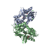
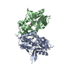
 Basic information
 Components
 Keywords
 Function and homology information
 Homo sapiens (human)
 X-RAY DIFFRACTION /
  Authors
 Citation
 Structure visualization
 Downloads & links
 Other downloads


 PDBj
PDBj


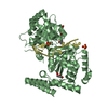
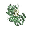
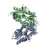
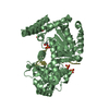
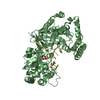
 Assembly
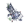
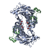


 Mass: 60.052 Da / Num. of mol.: 4 / Source method: obtained synthetically / Formula: C2H4O2
 Sample preparation
 Processing