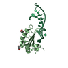
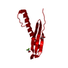
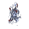
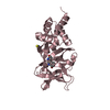
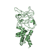
Entry Database : PDB / ID : 1mjsTitle MH2 domain of transcriptional factor SMAD3 SMAD 3 Keywords / Function / homology Function Domain/homology Component
/ / / / / / / / / / / / / / / / / / / / / / / / / / / / / / / / / / / / / / / / / / / / / / / / / / / / / / / / / / / / / / / / / / / / / / / / / / / / / / / / / / / / / / / / / / / / / / / / / / / / / / / / / / / / / / / / / / / / / / / / / / / / / / / / / / / / / Biological species Homo sapiens (human)Method / / / Resolution : 1.91 Å Authors Qin, B.Y. / Lam, S.S. / Correia, J.J. / Lin, K. Journal : Genes Dev. / Year : 2002Title : Smad3 allostery links TGF-beta receptor kinase activation to transcriptional controlAuthors : Qin, B.Y. / Lam, S.S. / Correia, J.J. / Lin, K. History Deposition Aug 28, 2002 Deposition site / Processing site Revision 1.0 Oct 16, 2002 Provider / Type Revision 1.1 Apr 28, 2008 Group Revision 1.2 Jul 13, 2011 Group Revision 1.3 Feb 14, 2024 Group / Database references / Category / chem_comp_bond / database_2Item / _database_2.pdbx_database_accessionRevision 1.4 Apr 3, 2024 Group / Category
Show all Show less
 Movie
Movie Controller


 Open data
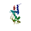
 Basic information
 Components
 Keywords
 Function and homology information
 Homo sapiens (human)
 X-RAY DIFFRACTION /
  Authors
 Citation
 Structure visualization
 Downloads & links
 Other downloads


 PDBj
PDBj


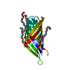
 Assembly
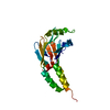

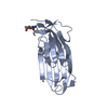
 Mass: 18.015 Da / Num. of mol.: 195 / Source method: isolated from a natural source / Formula: H2O
 Sample preparation
 / Beamline: X25 / Wavelength: 1.1 Å
 Processing