 Movie
Movie Controller

[English] 日本語
 Yorodumi
- PDB-1qjp: HIGH RESOLUTION STRUCTURE OF THE OUTER MEMBRANE PROTEIN A (OMPA) ... -
+ Open data

- Basic information
| Entry | Database: PDB / ID: 1qjp | ||||||
|---|---|---|---|---|---|---|---|
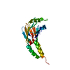
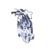
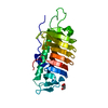
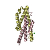
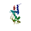
| Title | HIGH RESOLUTION STRUCTURE OF THE OUTER MEMBRANE PROTEIN A (OMPA) TRANSMEMBRANE DOMAIN | ||||||
 Components Components | OUTER MEMBRANE PROTEIN A | ||||||
 Keywords Keywords | OUTER MEMBRANE | ||||||
| Function / homology |  Function and homology information Function and homology information: / outer membrane protein complex / monoatomic ion transmembrane transporter activity / detection of virus / outer membrane / porin activity / pore complex / monoatomic ion transport / cell outer membrane / outer membrane-bounded periplasmic space ...: / outer membrane protein complex / monoatomic ion transmembrane transporter activity / detection of virus / outer membrane / porin activity / pore complex / monoatomic ion transport / cell outer membrane / outer membrane-bounded periplasmic space / monoatomic ion transmembrane transport / DNA damage response / symbiont entry into host cell / membrane / identical protein binding Similarity search - Function | ||||||
| Biological species |  | ||||||
| Method |  X-RAY DIFFRACTION / SYNCHROTRON / MOLECULAR REPLACEMENT / Resolution: 1.65 Å X-RAY DIFFRACTION / SYNCHROTRON / MOLECULAR REPLACEMENT / Resolution: 1.65 Å | ||||||
 Authors Authors | Pautsch, A. / Schulz, G.E. | ||||||
 Citation Citation | Journal: J.Mol.Biol. / Year: 2000 Title: High Resolution Structure of the Ompa Membrane Domain Authors: Pautsch, A. / Schulz, G.E. #1: Journal: Nat.Struct.Biol. / Year: 1998Title: Structure of the Outer Membrane Protein a Transmembrane Domain Authors: Pautsch, A. / Schulz, G.E. | ||||||
| History |
|
- Structure visualization
| Structure viewer | Molecule: MolmilJmol/JSmol |
|---|
- Downloads & links
-Download
| PDBx/mmCIF format | 1qjp.cif.gz | 79.2 KB | Display | PDBx/mmCIF format |
|---|---|---|---|---|
| PDB format | pdb1qjp.ent.gz | 58.9 KB | Display | PDB format |
| PDBx/mmJSON format | 1qjp.json.gz | Tree view | PDBx/mmJSON format | |
| Others |  Other downloads Other downloads |
-Validation report
| Arichive directory | https://data.pdbj.org/pub/pdb/validation_reports/qj/1qjpftp://data.pdbj.org/pub/pdb/validation_reports/qj/1qjp | HTTPS FTP |
|---|
-Related structure data
| Related structure data |  1bxwS S: Starting model for refinement |
|---|---|
| Similar structure data |

-Links
 PDBj
PDBj

- Assembly
| Deposited unit | 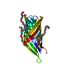
| ||||||||
|---|---|---|---|---|---|---|---|---|---|
| 1 |
| ||||||||
| Unit cell |
| ||||||||
| Details | BIOLOGICAL_UNIT: MONOMER |
-Components
| #1: Protein | Mass: 18795.719 Da / Num. of mol.: 1 / Fragment: TRANSMEMBRANE DOMAIN / Mutation: YES Source method: isolated from a genetically manipulated source Source: (gene. exp.) | ||||
|---|---|---|---|---|---|
| #2: Chemical | ChemComp-C8E / (   Mass: 306.438 Da / Num. of mol.: 6 / Source method: obtained synthetically / Formula: C16H34O5 / Comment: C8E, detergent*YM Mass: 306.438 Da / Num. of mol.: 6 / Source method: obtained synthetically / Formula: C16H34O5 / Comment: C8E, detergent*YM#3: Water | ChemComp-HOH / |  Mass: 18.015 Da / Num. of mol.: 64 / Source method: isolated from a natural source / Formula: H2O Mass: 18.015 Da / Num. of mol.: 64 / Source method: isolated from a natural source / Formula: H2OCompound details | F23L, Q34K AND K107Y WERE INTRODUCED | |
-Experimental details
-Experiment
| Experiment | Method: X-RAY DIFFRACTION / Number of used crystals: 1 |
|---|
- Sample preparation
| Crystal | Density Matthews: 3.5 Å3/Da / Density % sol: 65 % | |||||||||||||||||||||||||||||||||||
|---|---|---|---|---|---|---|---|---|---|---|---|---|---|---|---|---|---|---|---|---|---|---|---|---|---|---|---|---|---|---|---|---|---|---|---|---|
| Crystal grow | pH: 5 / Details: 10% PEG 8000, 10 % MPD, 25 MM KH2PO4 PH 5.1 | |||||||||||||||||||||||||||||||||||
| Crystal grow | *PLUS pH: 4.6 / Method: vapor diffusion, hanging drop | |||||||||||||||||||||||||||||||||||
| Components of the solutions | *PLUS
|
-Data collection
| Diffraction | Mean temperature: 100 K |
|---|---|
| Diffraction source | Source: SYNCHROTRON / Site: EMBL/DESY, HAMBURG  / Beamline: BW7B / Wavelength: 0.9 / Beamline: BW7B / Wavelength: 0.9 |
| Detector | Type: MARRESEARCH / Detector: IMAGE PLATE / Date: Apr 15, 1998 |
| Radiation | Protocol: SINGLE WAVELENGTH / Monochromatic (M) / Laue (L): M / Scattering type: x-ray |
| Radiation wavelength | Wavelength: 0.9 Å / Relative weight: 1 |
| Reflection | Resolution: 1.65→20 Å / Num. obs: 29702 / % possible obs: 95.4 % / Redundancy: 2.5 % / Biso Wilson estimate: 26 Å2 / Rsym value: 0.061 / Net I/σ(I): 13.7 |
| Reflection shell | Resolution: 1.65→1.75 Å / Redundancy: 2 % / Mean I/σ(I) obs: 3.2 / Rsym value: 0.307 / % possible all: 94.2 |
| Reflection | *PLUS Rmerge(I) obs: 0.061 |
| Reflection shell | *PLUS % possible obs: 94 % / Rmerge(I) obs: 0.31 |
- Processing
| Software |
| ||||||||||||||||||||||||||||||||||||||||||||||||||||||||||||||||||||||||||||||||||||
|---|---|---|---|---|---|---|---|---|---|---|---|---|---|---|---|---|---|---|---|---|---|---|---|---|---|---|---|---|---|---|---|---|---|---|---|---|---|---|---|---|---|---|---|---|---|---|---|---|---|---|---|---|---|---|---|---|---|---|---|---|---|---|---|---|---|---|---|---|---|---|---|---|---|---|---|---|---|---|---|---|---|---|---|---|---|
| Refinement | Method to determine structure: MOLECULAR REPLACEMENT Starting model: PDB ENTRY 1BXW Resolution: 1.65→12 Å / SU B: 0.60174 / SU ML: 0.02056 / Cross valid method: THROUGHOUT / σ(F): 0 / ESU R: 0.08 / ESU R Free: 0.07 Details: THE REGIONS Y18-T30, K64-G70 AND N146-N159 WERE DISORDERED AND ARE NOT INCLUDED IN THE MODEL
| ||||||||||||||||||||||||||||||||||||||||||||||||||||||||||||||||||||||||||||||||||||
| Displacement parameters | Biso mean: 44 Å2 | ||||||||||||||||||||||||||||||||||||||||||||||||||||||||||||||||||||||||||||||||||||
| Refinement step | Cycle: LAST / Resolution: 1.65→12 Å
| ||||||||||||||||||||||||||||||||||||||||||||||||||||||||||||||||||||||||||||||||||||
| Refine LS restraints |
| ||||||||||||||||||||||||||||||||||||||||||||||||||||||||||||||||||||||||||||||||||||
| Software | *PLUS Name: REFMAC / Classification: refinement | ||||||||||||||||||||||||||||||||||||||||||||||||||||||||||||||||||||||||||||||||||||
| Refine LS restraints | *PLUS
|
