 Movie
Movie Controller

[English] 日本語
 Yorodumi
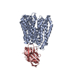
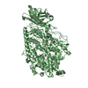
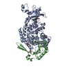
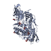
- PDB-6gs1: Crystal structure of peptide transporter DtpA-nanobody in MES buffer -
+ Open data

- Basic information
| Entry | Database: PDB / ID: 6gs1 | |||||||||
|---|---|---|---|---|---|---|---|---|---|---|
| Title | Crystal structure of peptide transporter DtpA-nanobody in MES buffer | |||||||||
 Components Components |
| |||||||||
 Keywords Keywords | MEMBRANE PROTEIN / peptide transporter / complex with nanobody / SLC15 family member | |||||||||
| Function / homology |  Function and homology information Function and homology informationdipeptide transmembrane transport / tripeptide transmembrane transport / tripeptide transmembrane transporter activity / peptide:proton symporter activity / dipeptide transmembrane transporter activity / protein transport / plasma membrane Similarity search - Function | |||||||||
| Biological species |   | |||||||||
| Method |  X-RAY DIFFRACTION / SYNCHROTRON / MOLECULAR REPLACEMENT / Resolution: 3.29 Å X-RAY DIFFRACTION / SYNCHROTRON / MOLECULAR REPLACEMENT / Resolution: 3.29 Å | |||||||||
 Authors Authors | Ural-Blimke, Y. / Flayhan, A. / Loew, C. / Quistgaard, E.M. | |||||||||
| Funding support |  Sweden, 2items Sweden, 2items
| |||||||||
 Citation Citation | Journal: J. Am. Chem. Soc. / Year: 2019 Title: Structure of Prototypic Peptide Transporter DtpA from E. coli in Complex with Valganciclovir Provides Insights into Drug Binding of Human PepT1. Authors: Ural-Blimke, Y. / Flayhan, A. / Strauss, J. / Rantos, V. / Bartels, K. / Nielsen, R. / Pardon, E. / Steyaert, J. / Kosinski, J. / Quistgaard, E.M. / Low, C. | |||||||||
| History |
|
- Structure visualization
| Structure viewer | Molecule: MolmilJmol/JSmol |
|---|
- Downloads & links
-Download
| PDBx/mmCIF format | 6gs1.cif.gz | 127 KB | Display | PDBx/mmCIF format |
|---|---|---|---|---|
| PDB format | pdb6gs1.ent.gz | 97.4 KB | Display | PDB format |
| PDBx/mmJSON format | 6gs1.json.gz | Tree view | PDBx/mmJSON format | |
| Others |  Other downloads Other downloads |
-Validation report
| Arichive directory | https://data.pdbj.org/pub/pdb/validation_reports/gs/6gs1ftp://data.pdbj.org/pub/pdb/validation_reports/gs/6gs1 | HTTPS FTP |
|---|
-Related structure data
| Related structure data |  6gs4C  6gs7C  4q65S S: Starting model for refinement C: citing same article ( |
|---|---|
| Similar structure data | |
| Other databases |
|

-Links
 PDBj
PDBj


- Assembly
| Deposited unit | 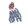
| ||||||||
|---|---|---|---|---|---|---|---|---|---|
| 1 |
| ||||||||
| Unit cell |
|
-Components
| #1: Protein | Mass: 54969.680 Da / Num. of mol.: 1 Source method: isolated from a genetically manipulated source Source: (gene. exp.) Strain: K12 / Gene: dtpA, tppB, ydgR, b1634, JW1626 / Production host: |
|---|---|
| #2: Antibody | Mass: 14400.683 Da / Num. of mol.: 1 Source method: isolated from a genetically manipulated source Source: (gene. exp.) |
| #3: Water | ChemComp-HOH /  Mass: 18.015 Da / Num. of mol.: 1 / Source method: isolated from a natural source / Formula: H2O Mass: 18.015 Da / Num. of mol.: 1 / Source method: isolated from a natural source / Formula: H2O |
| Has protein modification | Y |
-Experimental details
-Experiment
| Experiment | Method: X-RAY DIFFRACTION / Number of used crystals: 1 |
|---|
- Sample preparation
| Crystal | Density Matthews: 3.94 Å3/Da / Density % sol: 68.79 % |
|---|---|
| Crystal grow | Temperature: 292 K / Method: vapor diffusion, sitting drop / Details: PEG 600, magnesium chloride, MES buffer, pH 6.5 |
-Data collection
| Diffraction | Mean temperature: 100 K |
|---|---|
| Diffraction source | Source: SYNCHROTRON / Site: ESRF  / Beamline: MASSIF-1 / Wavelength: 0.966 Å / Beamline: MASSIF-1 / Wavelength: 0.966 Å |
| Detector | Type: DECTRIS PILATUS3 2M / Detector: PIXEL / Date: Feb 4, 2016 |
| Radiation | Protocol: SINGLE WAVELENGTH / Monochromatic (M) / Laue (L): M / Scattering type: x-ray |
| Radiation wavelength | Wavelength: 0.966 Å / Relative weight: 1 |
| Reflection | Resolution: 3.29→48.16 Å / Num. obs: 17262 / % possible obs: 99 % / Redundancy: 6.7 % / Rmerge(I) obs: 0.2673 / Net I/σ(I): 6.46 |
| Reflection shell | Resolution: 3.29→3.408 Å |
- Processing
| Software |
| |||||||||||||||||||||||||||||||||||||||||||||||||
|---|---|---|---|---|---|---|---|---|---|---|---|---|---|---|---|---|---|---|---|---|---|---|---|---|---|---|---|---|---|---|---|---|---|---|---|---|---|---|---|---|---|---|---|---|---|---|---|---|---|---|
| Refinement | Method to determine structure: MOLECULAR REPLACEMENT Starting model: 4q65 Resolution: 3.29→48.16 Å / SU ML: 0.53 / Cross valid method: FREE R-VALUE / σ(F): 1.36 / Phase error: 29.11
| |||||||||||||||||||||||||||||||||||||||||||||||||
| Solvent computation | Shrinkage radii: 0.9 Å / VDW probe radii: 1.11 Å | |||||||||||||||||||||||||||||||||||||||||||||||||
| Refinement step | Cycle: LAST / Resolution: 3.29→48.16 Å
| |||||||||||||||||||||||||||||||||||||||||||||||||
| Refine LS restraints |
| |||||||||||||||||||||||||||||||||||||||||||||||||
| LS refinement shell |
|
