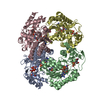
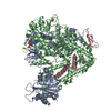
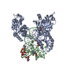
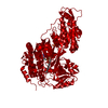
[English] 日本語
Yorodumi
- PDB-3zqj: Mycobacterium tuberculosis UvrA -

+
Open data


ID or keywords:

Loading...

-
Basic information

Entry
Database: PDB / ID: 3zqj
TitleMycobacterium tuberculosis UvrA
ComponentsUVRABC SYSTEM PROTEIN A
KeywordsDNA BINDING PROTEIN / NUCLEOTIDE EXCISION REPAIR
Function / homology
Function and homology information


negative regulation of strand invasion / excinuclease ABC activity / excinuclease repair complex / cell wall / SOS response / nucleotide-excision repair / DNA damage response / ATP hydrolysis activity / DNA binding / zinc ion binding ...negative regulation of strand invasion / excinuclease ABC activity / excinuclease repair complex / cell wall / SOS response / nucleotide-excision repair / DNA damage response / ATP hydrolysis activity / DNA binding / zinc ion binding / ATP binding / plasma membrane / cytosol / cytoplasm
Similarity search - Function
ABC transporter ATPase domain-like / ABC transporter ATPase like fold / ABC transporter ATPase like domain / UvrA, interaction domain / UvrA interaction domain / UvrABC system subunit A / UvrA DNA-binding domain / UvrA DNA-binding domain / Ribosomal protein L1/L10, rRNA-binding domain / Ribulose 1,5 Bisphosphate Carboxylase/Oxygenase ...ABC transporter ATPase domain-like / ABC transporter ATPase like fold / ABC transporter ATPase like domain / UvrA, interaction domain / UvrA interaction domain / UvrABC system subunit A / UvrA DNA-binding domain / UvrA DNA-binding domain / Ribosomal protein L1/L10, rRNA-binding domain / Ribulose 1,5 Bisphosphate Carboxylase/Oxygenase / Helicase, Ruva Protein; domain 3 / ABC transporter-like, conserved site / ABC transporters family signature. / ABC transporter / ABC transporter-like, ATP-binding domain / ATP-binding cassette, ABC transporter-type domain profile. / P-loop containing nucleotide triphosphate hydrolases / Up-down Bundle / P-loop containing nucleoside triphosphate hydrolase / Rossmann fold / 2-Layer Sandwich / Orthogonal Bundle / 3-Layer(aba) Sandwich / Mainly Alpha / Alpha Beta
Similarity search - Domain/homology
UvrABC system protein A / UvrABC system protein A
Similarity search - Component
Biological speciesMYCOBACTERIUM TUBERCULOSIS (bacteria)
MethodX-RAY DIFFRACTION / SYNCHROTRON / MOLECULAR REPLACEMENT / Resolution: 3.4 Å
AuthorsRossi, F. / Khanduja, J.S. / Bortoluzzi, A. / Houghton, J. / Sander, P. / Guthlein, C. / Davis, E.O. / Springer, B. / Bottger, E.C. / Relini, A. ...Rossi, F. / Khanduja, J.S. / Bortoluzzi, A. / Houghton, J. / Sander, P. / Guthlein, C. / Davis, E.O. / Springer, B. / Bottger, E.C. / Relini, A. / Penco, A. / Muniyappa, K. / Rizzi, M.
CitationJournal: Nucleic Acids Res. / Year: 2011
Title: The Biological and Structural Characterization of Mycobacterium Tuberculosis Uvra Provides Novel Insights Into its Mechanism of Action
Authors: Rossi, F. / Khanduja, J.S. / Bortoluzzi, A. / Houghton, J. / Sander, P. / Guthlein, C. / Davis, E.O. / Springer, B. / Bottger, E.C. / Relini, A. / Penco, A. / Muniyappa, K. / Rizzi, M.
History
DepositionJun 9, 2011Deposition site: PDBE / Processing site: PDBE
SupersessionJun 22, 2011ID: 2YGR
Revision 1.0Jun 22, 2011Provider: repository / Type: Initial release
Revision 1.1Sep 14, 2011Group: Database references
Revision 1.2Dec 20, 2023Group: Data collection / Database references ...Data collection / Database references / Derived calculations / Other / Refinement description
Category: chem_comp_atom / chem_comp_bond ...chem_comp_atom / chem_comp_bond / database_2 / pdbx_database_status / pdbx_initial_refinement_model / pdbx_struct_conn_angle / struct_conn / struct_site
Item: _database_2.pdbx_DOI / _database_2.pdbx_database_accession ..._database_2.pdbx_DOI / _database_2.pdbx_database_accession / _pdbx_database_status.status_code_sf / _pdbx_struct_conn_angle.ptnr1_auth_comp_id / _pdbx_struct_conn_angle.ptnr1_auth_seq_id / _pdbx_struct_conn_angle.ptnr1_label_atom_id / _pdbx_struct_conn_angle.ptnr1_label_comp_id / _pdbx_struct_conn_angle.ptnr1_label_seq_id / _pdbx_struct_conn_angle.ptnr2_auth_seq_id / _pdbx_struct_conn_angle.ptnr2_label_asym_id / _pdbx_struct_conn_angle.ptnr3_auth_comp_id / _pdbx_struct_conn_angle.ptnr3_auth_seq_id / _pdbx_struct_conn_angle.ptnr3_label_atom_id / _pdbx_struct_conn_angle.ptnr3_label_comp_id / _pdbx_struct_conn_angle.ptnr3_label_seq_id / _pdbx_struct_conn_angle.value / _struct_conn.pdbx_dist_value / _struct_conn.ptnr1_auth_comp_id / _struct_conn.ptnr1_auth_seq_id / _struct_conn.ptnr1_label_asym_id / _struct_conn.ptnr1_label_atom_id / _struct_conn.ptnr1_label_comp_id / _struct_conn.ptnr1_label_seq_id / _struct_conn.ptnr2_auth_comp_id / _struct_conn.ptnr2_auth_seq_id / _struct_conn.ptnr2_label_asym_id / _struct_conn.ptnr2_label_atom_id / _struct_conn.ptnr2_label_comp_id / _struct_conn.ptnr2_label_seq_id / _struct_site.pdbx_auth_asym_id / _struct_site.pdbx_auth_comp_id / _struct_site.pdbx_auth_seq_id

-
Structure visualization

Structure viewerMolecule:
MolmilJmol/JSmol

Downloads & links

-
Assembly

Deposited unit
A: UVRABC SYSTEM PROTEIN A
B: UVRABC SYSTEM PROTEIN A
C: UVRABC SYSTEM PROTEIN A
D: UVRABC SYSTEM PROTEIN A
E: UVRABC SYSTEM PROTEIN A
F: UVRABC SYSTEM PROTEIN A
hetero molecules


Theoretical massNumber of molelcules
Total (without water)653,75420
Polymers652,8386
Non-polymers91614
Water0
1
A: UVRABC SYSTEM PROTEIN A
hetero molecules


Theoretical massNumber of molelcules
Total (without water)109,0034
Polymers108,8061
Non-polymers1963
Water0
TypeNameSymmetry operationNumber
identity operation1_555x,y,z1
MethodPISA
2
B: UVRABC SYSTEM PROTEIN A
hetero molecules


Theoretical massNumber of molelcules
Total (without water)109,0034
Polymers108,8061
Non-polymers1963
Water0
TypeNameSymmetry operationNumber
identity operation1_555x,y,z1
MethodPISA
3
C: UVRABC SYSTEM PROTEIN A
hetero molecules


Theoretical massNumber of molelcules
Total (without water)109,0034
Polymers108,8061
Non-polymers1963
Water0
TypeNameSymmetry operationNumber
identity operation1_555x,y,z1
MethodPISA
4
D: UVRABC SYSTEM PROTEIN A
hetero molecules


Theoretical massNumber of molelcules
Total (without water)109,0034
Polymers108,8061
Non-polymers1963
Water0
TypeNameSymmetry operationNumber
identity operation1_555x,y,z1
MethodPISA
5
E: UVRABC SYSTEM PROTEIN A
hetero molecules


Theoretical massNumber of molelcules
Total (without water)108,9373
Polymers108,8061
Non-polymers1312
Water0
TypeNameSymmetry operationNumber
identity operation1_555x,y,z1
MethodPISA
6
F: UVRABC SYSTEM PROTEIN A


Theoretical massNumber of molelcules
Total (without water)108,8061
Polymers108,8061
Non-polymers00
Water0
TypeNameSymmetry operationNumber
identity operation1_555x,y,z1
MethodPISA
Unit cell
Length a, b, c (Å)258.227, 258.227, 204.554
Angle α, β, γ (deg.)90.00, 90.00, 120.00
Int Tables number154
Space group name H-MP3221
Noncrystallographic symmetry (NCS)NCS domain:
IDEns-IDDetails
11A
21B
31C
41D
51E
12A
22B
32C
42D
52E

NCS domain segments:
Dom-IDComponent-IDEns-IDRefine codeAuth asym-IDAuth seq-ID
1111A1 - 114
2111B1 - 114
3111C1 - 114
4111D1 - 114
5111E1 - 114
1121A463 - 999
2121B463 - 999
3121C463 - 999
4121D463 - 999
5121E463 - 999

NCS ensembles :
ID
1
2

-
Components

#1: Protein
UVRABC SYSTEM PROTEIN A / UVRA PROTEIN / EXCINUCLEASE ABC SUBUNIT A


Mass: 108806.398 Da / Num. of mol.: 6
Source method: isolated from a genetically manipulated source
Source: (gene. exp.) MYCOBACTERIUM TUBERCULOSIS (bacteria) / Production host: ESCHERICHIA COLI (E. coli) / Strain (production host): BL21(DE3) / References: UniProt: P63380, UniProt: P9WQK7*PLUS
#2: Chemical
ChemComp-ZN / ZINC ION


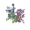
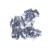
Mass: 65.409 Da / Num. of mol.: 14 / Source method: obtained synthetically / Formula: Zn

-
Experimental details

-
Experiment

ExperimentMethod: X-RAY DIFFRACTION / Number of used crystals: 1

-
Sample preparation

CrystalDensity Matthews: 3 Å3/Da / Density % sol: 60 % / Description: NONE
Crystal growpH: 7
Details: 1.5 M AMMONIUM SULFATE, 0.1 M HEPES PH 7.5, 1 MM YTTRIUM CHLORIDE.

-
Data collection

DiffractionMean temperature: 100 K
Diffraction sourceSource: SYNCHROTRON / Site: ESRF / Beamline: BM14 / Wavelength: 0.976
DetectorType: MARMOSAIC 225 mm CCD / Detector: CCD
RadiationProtocol: SINGLE WAVELENGTH / Monochromatic (M) / Laue (L): M / Scattering type: x-ray
Radiation wavelengthWavelength: 0.976 Å / Relative weight: 1
ReflectionResolution: 3.4→30 Å / Num. obs: 106710 / % possible obs: 98.9 % / Observed criterion σ(I): 0 / Redundancy: 2.4 % / Rmerge(I) obs: 0.09 / Net I/σ(I): 8.2
Reflection shellResolution: 3.4→3.58 Å / Redundancy: 2.4 % / Rmerge(I) obs: 0.43 / Mean I/σ(I) obs: 2 / % possible all: 100

-
Processing

Software
NameVersionClassification
REFMAC5.5.0044refinement
MOSFLMdata reduction
SCALAdata scaling
PHASERphasing
RefinementMethod to determine structure: MOLECULAR REPLACEMENT
Starting model: PDB ENTRY 2R6F
Resolution: 3.4→30 Å / Cor.coef. Fo:Fc: 0.879 / Cor.coef. Fo:Fc free: 0.801 / SU B: 33.705 / SU ML: 0.523 / Cross valid method: THROUGHOUT / ESU R Free: 0.682 / Stereochemistry target values: MAXIMUM LIKELIHOOD
Details: HYDROGENS HAVE BEEN ADDED IN THE RIDING POSITIONS.DISORDERED REGIONS WERE OMITTED FROM THE MODEL.
RfactorNum. reflection% reflectionSelection details
Rfree0.32422 1058 1 %RANDOM
Rwork0.27286 ---
obs0.27336 105162 100 %-
Solvent computationIon probe radii: 0.8 Å / Shrinkage radii: 0.8 Å / VDW probe radii: 1.4 Å / Solvent model: BABINET MODEL WITH MASK
Displacement parametersBiso mean: 63.328 Å2
Baniso -1Baniso -2Baniso -3
1-2.17 Å21.09 Å20 Å2
2--2.17 Å20 Å2
3----3.26 Å2
Refinement stepCycle: LAST / Resolution: 3.4→30 Å
ProteinNucleic acidLigandSolventTotal
Num. atoms39984 0 14 0 39998
Refine LS restraints
Refine-IDTypeDev idealDev ideal targetNumber
X-RAY DIFFRACTIONr_bond_refined_d0.0160.02240672
X-RAY DIFFRACTIONr_bond_other_d
X-RAY DIFFRACTIONr_angle_refined_deg1.7611.97555102
X-RAY DIFFRACTIONr_angle_other_deg
X-RAY DIFFRACTIONr_dihedral_angle_1_deg8.18555169
X-RAY DIFFRACTIONr_dihedral_angle_2_deg36.19623.0361841
X-RAY DIFFRACTIONr_dihedral_angle_3_deg24156875
X-RAY DIFFRACTIONr_dihedral_angle_4_deg19.59215422
X-RAY DIFFRACTIONr_chiral_restr0.1150.26287
X-RAY DIFFRACTIONr_gen_planes_refined0.0080.02130878
X-RAY DIFFRACTIONr_gen_planes_other
X-RAY DIFFRACTIONr_nbd_refined
X-RAY DIFFRACTIONr_nbd_other
X-RAY DIFFRACTIONr_nbtor_refined
X-RAY DIFFRACTIONr_nbtor_other
X-RAY DIFFRACTIONr_xyhbond_nbd_refined
X-RAY DIFFRACTIONr_xyhbond_nbd_other
X-RAY DIFFRACTIONr_metal_ion_refined
X-RAY DIFFRACTIONr_metal_ion_other
X-RAY DIFFRACTIONr_symmetry_vdw_refined
X-RAY DIFFRACTIONr_symmetry_vdw_other
X-RAY DIFFRACTIONr_symmetry_hbond_refined
X-RAY DIFFRACTIONr_symmetry_hbond_other
X-RAY DIFFRACTIONr_symmetry_metal_ion_refined
X-RAY DIFFRACTIONr_symmetry_metal_ion_other
X-RAY DIFFRACTIONr_mcbond_it0.7911.525719
X-RAY DIFFRACTIONr_mcbond_other
X-RAY DIFFRACTIONr_mcangle_it1.552241392
X-RAY DIFFRACTIONr_mcangle_other
X-RAY DIFFRACTIONr_scbond_it1.954314953
X-RAY DIFFRACTIONr_scbond_other
X-RAY DIFFRACTIONr_scangle_it3.6144.513710
X-RAY DIFFRACTIONr_scangle_other
X-RAY DIFFRACTIONr_long_range_B_refined
X-RAY DIFFRACTIONr_long_range_B_other
X-RAY DIFFRACTIONr_rigid_bond_restr
X-RAY DIFFRACTIONr_sphericity_free
X-RAY DIFFRACTIONr_sphericity_bonded
Refine LS restraints NCS

Refine-ID: X-RAY DIFFRACTION

Ens-IDDom-IDAuth asym-IDNumberTypeRms dev position (Å)Weight position
11A885tight positional0.050.05
12B885tight positional0.050.05
13C885tight positional0.060.05
14D885tight positional0.050.05
15E885tight positional0.060.05
21A3737tight positional0.050.05
22B3737tight positional0.050.05
23C3737tight positional0.050.05
24D3737tight positional0.050.05
25E3737tight positional0.050.05
11A885tight thermal0.070.5
12B885tight thermal0.070.5
13C885tight thermal0.080.5
14D885tight thermal0.080.5
15E885tight thermal0.080.5
21A3737tight thermal0.070.5
22B3737tight thermal0.070.5
23C3737tight thermal0.070.5
24D3737tight thermal0.080.5
25E3737tight thermal0.070.5
LS refinement shellResolution: 3.4→3.487 Å / Total num. of bins used: 20
RfactorNum. reflection% reflection
Rfree0.415 74 -
Rwork0.345 7731 -
obs--100 %

+
About Yorodumi

-
News

-
Feb 9, 2022. New format data for meta-information of EMDB entries

New format data for meta-information of EMDB entries

  • Version 3 of the EMDB header file is now the official format.
  • The previous official version 1.9 will be removed from the archive.

Related info.:EMDB header

External links:wwPDB to switch to version 3 of the EMDB data model

-
Aug 12, 2020. Covid-19 info

Covid-19 info

URL: https://pdbj.org/emnavi/covid19.php

New page: Covid-19 featured information page in EM Navigator.

Related info.:Covid-19 info / Mar 5, 2020. Novel coronavirus structure data

+
Mar 5, 2020. Novel coronavirus structure data

Novel coronavirus structure data

Related info.:Yorodumi Speices / Aug 12, 2020. Covid-19 info

External links:COVID-19 featured content - PDBj / Molecule of the Month (242):Coronavirus Proteases

+
Jan 31, 2019. EMDB accession codes are about to change! (news from PDBe EMDB page)

EMDB accession codes are about to change! (news from PDBe EMDB page)

  • The allocation of 4 digits for EMDB accession codes will soon come to an end. Whilst these codes will remain in use, new EMDB accession codes will include an additional digit and will expand incrementally as the available range of codes is exhausted. The current 4-digit format prefixed with “EMD-” (i.e. EMD-XXXX) will advance to a 5-digit format (i.e. EMD-XXXXX), and so on. It is currently estimated that the 4-digit codes will be depleted around Spring 2019, at which point the 5-digit format will come into force.
  • The EM Navigator/Yorodumi systems omit the EMD- prefix.

Related info.:Q: What is EMD? / ID/Accession-code notation in Yorodumi/EM Navigator

External links:EMDB Accession Codes are Changing Soon! / Contact to PDBj

+
Jul 12, 2017. Major update of PDB

Major update of PDB

  • wwPDB released updated PDB data conforming to the new PDBx/mmCIF dictionary.
  • This is a major update changing the version number from 4 to 5, and with Remediation, in which all the entries are updated.
  • In this update, many items about electron microscopy experimental information are reorganized (e.g. em_software).
  • Now, EM Navigator and Yorodumi are based on the updated data.

External links:wwPDB Remediation / Enriched Model Files Conforming to OneDep Data Standards Now Available in the PDB FTP Archive

-
Yorodumi

Thousand views of thousand structures

  • Yorodumi is a browser for structure data from EMDB, PDB, SASBDB, etc.
  • This page is also the successor to EM Navigator detail page, and also detail information page/front-end page for Omokage search.
  • The word "yorodu" (or yorozu) is an old Japanese word meaning "ten thousand". "mi" (miru) is to see.

Related info.:EMDB / PDB / SASBDB / Comparison of 3 databanks / Yorodumi Search / Aug 31, 2016. New EM Navigator & Yorodumi / Yorodumi Papers / Jmol/JSmol / Function and homology information / Changes in new EM Navigator and Yorodumi

Read more